Neues aus der Sportmedizin
Curathleticum
Blog
Erfahren Sie hier mehr zu medizinischen Erkenntnissen
und Methoden aus dem Spitzensport:
Im Curathleticum Blog veröffentliche ich regelmäßig
Auszüge aus meinen wissenschaftlichen Arbeiten
sowie Informationen zu interessanten Veranstaltungen,
Vorträgen und Events aus meinem Netzwerk.
DECEMBER 7, 2021
Return to play after treating acute muscle injuries in elite football players with radial extracorporeal shock wave therapy
Publikation in Journal of Orthopedic Surgery and Research (James P. M. Morgan¹, Mario Hamm², Christoph Schmitz¹* and Matthias H. Brem³,⁴)
Abstract
Background To compare lay-off times achieved by treating acute muscle injuries in elite football players with a multimodal
therapy approach that includes a specific protocol of almost daily radial extracorporeal shock wave therapy
(rESWT) with corresponding data reported in the literature.
Methods We performed a retrospective analysis of treatments and recovery times of muscle injuries suffered by
the players of an elite football team competing in the first/second German Bundesliga during one of the previous
seasons.
Results A total of 20 acute muscle injuries were diagnosed and treated in the aforementioned season, of which
eight (40%) were diagnosed as Type 1a/muscular tightness injuries, five (25%) as Type 2b/muscle strain injuries, four
(20%) as Type 3a/partial muscle tear injuries and three (15%) as contusions. All injuries were treated with the previously
mentioned multimodal therapy approach. Compared with data reported by Ekstrand et al. (Br J Sports Med
47:769–774, 2013), lay-off times (median/mean) were shortened by 54% and 58%, respectively, in the case of Type
1a injuries, by 50% and 55%, respectively, in the case of Type 2b injuries as well as by 8% and 21%, respectively, in the
case of Type 3a injuries. No adverse reactions were observed.
Conclusions Overall, the multimodal therapy approach investigated in this study is a safe and effective treatment
approach for treating Type 1a and 2b acute muscle injuries amongst elite football players and may help to prevent
more severe, structural muscle injuries.
Keywords Acute muscle injury, Athletes, Extracorporeal shock wave therapy, Rehabilitation, Return-to-play
Background
Muscle injuries are the most common injury in football [1, 2]. Non-structural injuries can contribute to over 50% of missed training sessions and participation in sporting activities and may worsen to become structural injuries if not appropriately managed [3]. According to Müller-Wohlfahrt et al. [4], muscle injuries can be classified into indirect muscle injuries (comprising functional and structural injuries) and direct injuries (contusion, laceration) (Table 1). Some authors proposed to classify functional injuries as ultrastructural injuries [5]. Further classification of structural muscle injuries involves definition of the exact site of the lesion within the musculotendinous unit ranging from proximal (P), middle (M) and distal (D) [3].
Recent approaches to improve therapy for acute muscle injuries have mainly focused on Type 3b injuries (structural injuries involving significant muscle tears) [6, 7]. In contrast, the treatment of Type 1a and 2b functional/ultrastructural muscle injuries as well as of Type 3a structural muscle injuries (smaller partial muscle tears) has largely been neglected in the academic literature during the last few decades, although these injuries can also cause considerable lay-off times of two weeks or longer [2, 8]. In the guidelines for muscle injuries outlined by the Italian Society of Muscles, Ligaments and Tendons, Maffulli et al. [8] recommended a multimodal therapy approach comprising RICE (rest, ice, compression, elevation), optimized loading, manual therapy, functional compression bandages, low-level laser therapy, pulsed ultrasound therapy, electroanalgesia, training and functional rehabilitation, without reference to specific evidence in the literature or modifying the treatment plan to cater for different types of injury severity.
During the past few years, studies using animal subjects with acute muscle injuries and in vitro studies have shown that radial and focused extracorporeal shock wave therapy (rESWT, fESWT) may be of benefit in treating acute muscle injuries [9, 10]. This form of treatment has already become well-established in successfully managing other pathologies of the musculoskeletal system, such as in the treatment of tendinopathies and fracture malunions [11, 12]. Radial and focused extracorporeal shock waves (rESWs, fESWs) are single acoustic impulses which have an initial high positive peak pressure between 10 and 100 megapascals that is reached in less than one microsecond, followed by a low tensile amplitude of a few µs duration that can generate cavitation, and a short life cycle of approximately 10–20 μs [13, 14]. Due to these characteristics rESWs and fESWs fundamentally differ from therapeutic ultrasound. Focused ESWs differ from rESWs in terms of how the shock waves are generated. Focused ESWs also differ in terms of their physical characteristics and with regards to the penetration depth of the shock waves into the tissue [11, 14,15,16]. Studies on rESWT/fESWT for the treatment of acute muscle injuries in elite football players (or other sportspeople) have not yet been published.
Based on anecdotal evidence we introduced rESWT (and to a much lesser extent, also fESWT) into our multimodal therapy approach for treating acute muscle injuries amongst elite football players (first/second German Bundesliga). Other components of this multimodal therapy approach included cryotherapy, compression, manual therapy, resistance/weight-training and a progressive physiotherapy exercise programme (in line with Maffulli et al. [8]).
In this study, we report our experience from the first entire season during which rESWT was applied. Our retrospective analysis was performed under the hypothesis that by integrating rESWT into our multimodal therapy approach, lay-off times may be shortened and a reduction in re-injury rates following acute functional/ultrastructural and structural muscle injuries amongst elite football players may be achieved.
Methods
Participants
We performed a retrospective analysis of treatments and time courses of all muscle injuries suffered by the players of an elite football team during one of the previous seasons (first/second German Bundesliga). This study was approved by the football club whose players were included in the study, and the local ethics board of Friedrich-Alexander University Erlangen-Nuremberg (Erlangen, Germany) (registration number 387_17 Bc). Written informed consent was obtained from all players for using their data in this study. CS did not visit the football club during diagnosis and treatment sessions and only had access to fully anonymous data.
All players were male and aged between 18 and 35 years old. All other details (including the dates of injury, side of injury, the player’s position, whether an injured player was left-footed or right-footed, and whether an injury involved a player’s support leg or kicking leg) are subject to confidentiality in order to protect the identity of each player within this study.
Diagnosis and definition of acute muscle injuries
The diagnosis of the acute muscle injuries within this study followed international guidelines and widely accepted clinical practice [8]. In summary, the diagnosis of functional/ultrastructural injuries (Type 1a and Type 2b injuries, respectively) was based on clinical examination. Specifically, functional tests such as hopping on one leg were used to provoke pain as well as to test the player’s readiness to commence (running) training; muscle length testing (stretch tests) and comparison with the non-injured limb, manual muscle strength testing and comparison with the non-injured limb, palpation of the injury site and comparison with the non-injured limb and a comprehensive subjective examination was undertaken. Pain was recorded using the Visual Analogue Scale (VAS) during the clinical examination. The subjective examination included noting the mechanism of injury and the exact injury location as well as noting the nature or quality of the pain being experienced (tension, tightness, sharp pain, burning, etc.). The diagnosis of Type 2b, 3a and 3b injuries, respectively, was also performed by using the previously described clinical examination methods. In addition, ultrasound scans and magnetic resonance imaging (MRI) was used to differentiate between injury severity amongst these groups of injuries. Contusions were diagnosed based on the patient’s history, clinical examination and ultrasound examination.
Multimodal therapy approach for treating acute muscle injuries
All acute muscle injuries were treated with a customized multimodal therapy approach comprising cryotherapy, compression, manual therapy, resistance/weight-training, a progressive physiotherapy exercise programme and ESWT (an example is provided in Table 2).
In 19 out of 20 cases rESWT was performed, using a Swiss DolorClast device (Electro Medical Systems, Nyon, Switzerland) equipped with an EvoBlue handpiece and 36-mm applicator. Radial ESWs were applied at 20 Hertz (Hz). In the majority of cases rESWT was performed on a daily basis. The energy density of the rESWs was individually adjusted—so that the player reported some discomfort but did not experience pain during treatment, resulting in an air pressure of between 1.0 and 3.4 bar. A single treatment session consisted of between 6.000 and 12.000 rESWs being applied. Individual rESWT protocols are displayed in Fig. 1. The decision to incorporate rESWT protocols into our treatment plan for muscle injuries was based on our promising earlier clinical experience and observations as well as positive subjective player information and feedback indicating that faster recovery times may be achievable in comparison to treatments of muscle injuries without the use of rESWT.
Protocols of radial extracorporeal shock wave therapy (rESWT) (A–S) or focused extracorporeal shock wave therapy (fESWT) (T) of all acute muscle injuries type 1a (A–H), 2b (I-M), 3a (N–Q) and contusions (R–T) suffered by the players of an elite football team during one of the previous seasons (first/second German Bundesliga), arranged in order of increasing lay-off times. All rESWT treatments were performed with a Swiss DolorClast device (Electro Medical Systems, Nyon, Switzerland) equipped with EvoBlue handpiece and 36-mm applicator; radial extracorporeal shock waves (rESWs) were applied at 20 Hz. Air pressure data are marked by red dots (between 2 and 3.5 bar) and the number of rESWs per treatment session by blue dots (between 6.000 and 12,000 per treatment session). The fESWT treatment shown in T was performed with a Swiss PiezoClast (Electro Medical Systems) and 15-mm gel pad; focused extracorporeal shock waves (fESWs) were applied at 8 Hz. Energy level data are marked by red dots (Level 10) and the number of fESWs per treatment session by blue dots (between 2.500 and 3.000 per treatment). In each case, Day 0 was the day of injury. Every pair of red and blue dots indicates a single treatment session. When the player’s status was 4 on the day of the last treatment (explained in Fig. 2), return to play was achieved on this day. In contrast, when the player’s status was 3 on the day of the last treatment (explained in Fig. 2), return to play was achieved on the following day. Delays in starting with rESWT were due to away games and traveling
One case (contusion of the gluteus maximus muscle) was treated with fESWT, using a Swiss PiezoClast device (Electro Medical Systems) and 15-mm gel pad. Focused ESWs were applied at 8 Hz; the positive energy density of the fESWs was 0.13 milliJoule per square millimeter (energy level 10). A single treatment session consisted of between 2.500 and 3.000 fESWs being applied. The corresponding fESWT protocol is shown in Fig. 1Q.
In the vast majority of cases rESWT/fESWT was commenced on the day of the injury (9/20 = 45%), the day after the injury (4/20 = 25%) or two days after the injury (3/20 = 15%). Delays in starting with rESWT/fESWT were due to away games or traveling and as a result not having immediate access to rESWT/fESWT.
There were no contraindications to rESWT/fESWT amongst the group of players that were treated. Contraindications to rESWT include as follows: local steroid injections during the last six weeks before rESWT/fESWT, infection or tumor at the site of rESWT/fESWT application, serious blood dyscrasia, blood-clotting disorders (including local thrombosis) and treatment with oral anticoagulants.
Return to sport
“Return-to-sport” status was defined as being once the player was able to fully participate in regular team training including contact training and was fully available for selection for matches in the first/second Bundesliga.
Results
A total of 20 acute muscle injuries occurred during the investigated season and were treated with the aforementioned approach, of which eight (40%) were diagnosed as Type 1a injuries, five (25%) as Type 2b injuries, four (20%) as Type 3a injuries and three (15%) as contusions. Accordingly, 13/17 = 76% of the injuries that were not contusions were functional/ultrastructural injuries (Types 1a and 2b), and 4/17 = 24% were structural injuries (Type 3a). There were no Type 3b or Type 4 injuries suffered during the investigated season.
Amongst the 17 injuries that were not classified as contusions, one (6%) occurred in the muscles in the anterior compartment of the thigh, four (24%) in the muscles in the medial compartment of the thigh, six (35%) in the posterior compartment of the thigh, and six (35%) in the posterior compartment of the leg.
Injuries occurred during the entire season. There was no difference in the number of injuries suffered at the beginning/first half of the season compared to the injuries suffered in the second half of the season after the winter break. All of the structural injuries (Type 3a) occurred several months after the start of the season. The re-injury rate (defined as being an injury involving the same muscle and having the same severity as the initial injury occurring within two months after a player’s return-to-sport following the initial injury [2]) was 1/8 = 12.5% for Type 1a injuries, and was zero in the case of Type 2b, 3a injuries and contusions, respectively. None of the Type 2b injuries that were diagnosed occurred following a previously suffered Type 1a injury during the investigated season, and none of the Type 3a injuries occurred following a previously suffered Type 1a or 2b injury during the investigated season.
Return-to-play was achieved after 3/3.3/2–6 days (median/mean/range), respectively, following Type 1a injuries, after 4/6.2/3–14 days, respectively, following Type 2b injuries, after 12/13/7–22 days, respectively, following Type 3a injuries and after 4/4/4 days, respectively, following contusions (Fig. 2).
The MRI pictures of the lower leg of a player who was diagnosed with a minor partial muscle tear (Type 3a) of the soleus muscle are shown in Fig. 3. In this case, rESWT was performed on days 4, 5, 7, 8, 12, 13, 15, 16 and 18–20, respectively, following injury (Figs. 1Q and 2Q). The detailed treatment protocol of this case is summarized in Table 2. Return-to-play was achieved on day 22 post-injury.
No adverse reactions to treatment were observed, except occasional, temporary reddening of the skin at the treatment site that disappeared within 24 h following rESWT.
All of the players who were treated for muscle injuries complied fully with the treatment protocol incorporating rESWT (or fESWT in one case). No player declined being treated with rESWT (or fESWT in one case) neither during the initial treatment nor during the follow-up treatments. Hence, by comparing the use of our treatment protocol for muscle injuries with other treatment protocols and the differences in lay-off times, respectively, one can conclude that non-compliance did not affect the treatment effect of our retrospective analysis.
Discussion
This is the first report concerning the course of recovery times following acute muscle injuries suffered by the players of an elite football team (competing in the first/second German Bundesliga) during an entire season that incorporated rESWT/fESWT into the treatment protocols of these injuries. A number of relevant conclusions can be drawn from these results (with reference to rESWT because fESWT was only used in one single case).
Safety of integrating rESWT into a multimodal therapy approach for treating acute muscle injuries
Integrating rESWT into a multimodal therapy approach for treating acute muscle injuries as outlined in this study is safe. There were no adverse reactions or complications to treatment observed apart from occasional, temporary reddening of the skin at the treatment site that disappeared within 24 h following rESWT. Moreover, there were no observed cases of local hematoma following the application of rESWs—as has been previously reported in the literature following the application of rESWs for the treatment of the gluteus maximus muscle with a rESWT device that differed from the one that was used in this study [17]. In addition, none of the players that were treated suffered from myositis ossificans (MO)—a proliferative mesenchymal response following soft tissue trauma that causes localized ossification [18]. This is important to note because, at first glance, exposure of injured muscles to rESWs could be considered inappropriate physiotherapy [19]. Walczak et al. [18] hypothesized (with reference to a study by Medici et al. [20]) that the development of MO may depend on a process called endothelial-mesenchymal transition. According to this hypothesis, skeletal muscle injury may induce a local inflammatory cascade, which leads to the release of bone morphogenetic protein (BMP)-2, BMP-4 and transforming growth factor (TGF). These cytokines may act on vascular endothelial cells and induce endothelial-mesenchymal transition. As a result, endothelial-derived mesenchymal stem cells may differentiate into osteoblasts and chondrocytes when exposed to an inflammatory-rich environment [18]. Accordingly, it is important to note that exposure of tissue to rESWs/fESWs (or, more specifically, exposure to fESWs; because related studies on rESWs have not yet been published) can increase local concentrations of BMP-2, BMP-4 and TGF-ß1 [21, 22]. If this is the case it would clearly be an argument against using rESWT/fESWT in the treatment of acute muscle injuries. However, it is important to keep in mind that the aforementioned hypothesis of endothelial-mesenchymal transition in the pathophysiology of MO is based on observations of a rare disease called fibrodysplasia ossificans progressiva [20] rather than on traumatic MO. A number of studies on human heterotopic ossification and related mouse models have demonstrated that bone marrow-derived osteoblast progenitor cells in circulating blood may contribute to the formation of heterotopic bone [23,24,25]. Moreover, a recent study involving a parabiosis model (i.e., two mice with shared circulation, in which the cells of one mouse can be labeled and detected in the other mouse when released into the bloodstream) has demonstrated that these bone marrow-derived osteoblast progenitor cells do not contribute to early stage development of heterotopic ossification [26]. The authors of this study argued that a timely intervention that influences the molecular and cellular processes that contribute to the recruitment and/or differentiation of the circulating cell populations that participate in heterotopic ossification may block the initiation of the latter and ultimately prevent the transition to definitive bone [26]. We believe that this may be achieved by the very early treatment of acute muscle injuries with rESWT, as performed in this study. Further studies on animal models are required to test this hypothesis. It is worth noting that in this study commencing rESWT
immediately following Type 1a and 2b injuries did not lead to inferior results when compared with starting treatment with rESWT on days 1, 3 or 4 post-injury (delay in commencing treatment was due to some injuries being suffered during away games that required traveling after the games and as a result there being no immediate access to rESWT/fESWT).
Shortening of lay-off times by integrating rESWT into a multimodal therapy approach for treating acute muscle injuries
Integrating rESWT into a multimodal therapy approach for the treatment of acute muscle injuries as performed in this study may shorten lay-off times for elite football players (as well as for other sportspeople) compared to other therapy approaches. This conclusion is based on a comparison of the results of this study with data that were obtained prospectively from 31 European elite male football teams during the 2011/2012 season (Table 3) [2].
A direct comparison with the data reported by Ekstrand et al. [2] shows that the median/mean lay-off times in this study were shortened by 54%/58%, respectively, in the case of Type 1a injuries, by 50%/55%, respectively, in the case of Type 2b injuries, and by 8%/21%, respectively, in the case of Type 3a injuries. Arguably, the Type 3a structural injury shown in Fig. 3 could also be considered to be a more severe Type 3b injury rather than a Type 3a injury which would mean that the median/mean injury lay-off times in this study for Type 3a injuries would then be further shortened by 23%/37%, respectively, compared to the data reported by Ekstrand et al. [2]. Furthermore, in recently published guidelines for muscle injuries, Maffulli et al. [8] consider normal lay-off times to be 5–15 days for functional/ultrastructural injuries, 15–18 days for Type 3a injuries and 25–35 days for Type 3b injuries. The lay-off times reported in this study were substantially shorter than the aforementioned typical lay-off times noted by Maffulli et al. [8]. At this stage, it is difficult to ascertain to which extent the application of rESWs actually contributed to the shorter lay-off times in this study compared to the data reported by Ekstrand et al. [2]. This is partly due to the fact that no treatment protocols were outlined in the latter study. Nevertheless, several factors strengthen the argumentation that the application of rESWs may be helpful as an adjunct form of therapy for reducing lay-off times following muscle injuries. Specifically: (1) the study by Ekstrand et al. [2] was based on data that was obtained from European male elite football teams and, therefore, from a population that is very similar to the population investigated in this study; (2) the key molecular and cellular mechanisms of action that rESWs/fESWs have on muscles (outlined below) were not yet known in 2011/2012; (3) Maffulli et al. [8] did not mention rESWT/fESWT in their guidelines for muscle injuries; and (4) our multimodal therapy approach (excluding rESWT/fESWT) is in line with the guidelines published by Maffulli et al. [8]. With this in mind, it is reasonable to assume that the therapy approaches used in 2011/2012 by the teams who were investigated by Ekstrand et al. [2] were broadly comparable to the multimodal therapy approach (excluding rESWT/fESWT) used in this study. Hence the application of rESWs may have substantially contributed to the shorter lay-off times found in this study compared to the data reported by Ekstrand et al. [2]. Further studies are required to test this hypothesis.
Reduction in total re-injury rates by integrating rESWT into a multimodal therapy approach for treating acute muscle injuries
the total re-injury rates of 1/13 (8%) observed amongst functional/ultrastructural muscle injuries and 0/4 (0%) observed amongst the structural muscle injuries in this study were lower than corresponding data reported by Ekstrand et al. [2] (12% was observed amongst functional/ultrastructural injuries and 13% observed amongst structural injuries, respectively). Based on these results we argue that the integration of rESWT into a multimodal therapy approach for the treatment of acute muscle injuries as outlined in this study may not only help to reduce lay-off times but may also help in the prevention of muscle re-injury amongst athletes. However, due to the low number of structural muscle injuries included in this study, further studies are required to test this hypothesis amongst a larger sample group.
In addition, no Type 2b injuries occurred in this study following a Type 1a injury and no structural injury occurred following a functional/ultrastructural injury. In contrast, Ekstrand et al. [2] reported in their study, that 5% of the initial functional/ultrastructural injuries developed into secondary structural injuries within two months of the primary injury. However, they did not report how many Type 2b injuries occurred following an earlier Type 1a injury during the investigated season, nor did they comment on how many structural injuries occurred following previously suffered functional/ultrastructural injuries during the entire investigated season (i.e., not only including the initial two months, or “re-injury” period following the primary injury). It is important to note that in this study, the number of functional/ultrastructural compared to structural acute muscle injuries was 13 as opposed to four. Hence there were considerably more functional/ultrastructural injuries included in this study, but this number was 130 as opposed to 263 (i.e., more structural injuries) in the study by Ekstrand et al. [2]. Of course, many other factors may have contributed to this finding—such as differences in the management of training load upon returning to team training, and other prevention strategies used such as optimizing nutrition and using screening protocols for detecting early warning signs that may allow sport medical staff to help “forecast” an impending muscle injury [27,28,29]. In a recent study regular eccentric lengthening of the muscles in the lower limbs as performed in the practice of Salah (a religious prayer amongst Muslims) was found to reduce the number of structural muscle injuries amongst professional Russian football players when compared with a control group [30]. Exercises involving the eccentric lengthening of muscles were also used as a component of the physiotherapy exercises integrated into the multimodal therapy approach used in this study. Furthermore, decreasing delayed onset muscle soreness by whole body vibration could also contribute to reduced muscle injury risk in elite football players, as recently shown in elite hockey players [31]. However, one cannot rule out that the integration of rESWT into a multimodal therapy approach for the treatment of acute muscle injuries as performed in this study may also contribute to the prevention of structural muscle injuries in athletes.
Limitations
This study is an audit of retrospectively collected data, and therefore has a number of inherent limitations. Firstly, there was no control group. The reason for this was that the evidence that was available at the time when we decided to integrate rESWT/fESWT into our therapy approach was not sufficient enough to gain approval from the club for a possible prospective study with a control group. Paradoxically, the promising results of this study (in particular the comparison to the data reported by Ekstrand et al. [2]) may now also cause difficulties in gaining approval from the same or another club for a corresponding prospective study with a control group, as the clubs‘ interests obviously lie in reducing lay-off times following injuries to a minimum. Moreover, gaining approval for a corresponding randomized controlled trial (RCT) from the players themselves may also prove to be more difficult following the initial good results obtained from using rESWT/fESWT in this study. Perhaps it is more realistic to expect that future RCTs be performed on recreational athletes, as in the case of one current study that relates to the treatment of acute Type 3b hamstring muscle injuries [16].
Secondly, with the exception of one case (contusion) only rESWT was investigated in this study. One may argue that fESWT may also be effective in the management of acute muscle injuries amongst athletes, but obviously there are not enough data to support this hypothesis. Our decision to focus on rESWT rather than on fESWT was based on the fact that the data relating to treatment of other pathologies of the musculoskeletal system (tendinopathies and fracture malunions of superficial bones) do not support the hypothesis that fESWT is superior to rESWT [11, 12]. In addition, the use of fESWT is restricted to physicians in many countries (as is the case in Germany where physiotherapists and chiropractors who have been trained in Germany are not legally entitled to use fESWT). Moreover, the International Society for Medical Shockwave Treatment (ISMST) has recommended that only a qualified physician (certified by National or International Societies) may use fESWT in their latest Consensus Statement regarding ESWT indications and contraindications [32]. However, in many elite football clubs (and training centers involving other sporting codes) a medical doctor may not be available to perform treatment on a daily basis. This is also the reason why a RCT on acute Type 3b hamstring muscle injuries currently being undertaken is based on rESWT rather than on fESWT [16].
Thirdly, only one rESWT protocol was applied in this study, and this protocol differed considerably from other published rESWT protocols used in the treatments of tendinopathies [11]. Specifically, there were differences regarding the timing with regards to commencing rESWT (immediately after the injury in this study compared with starting treatment six or more months after initial diagnosis and following unsuccessful treatment involving other conservative modalities [33,34,35]), differences in the time interval between treatment sessions (in most cases daily treatment sessions were used in this study as compared to one treatment session per week used in other studies [11]) and differences in the number of rESWs applied per treatment session (6.000–12.000 in this study as opposed to treatments averaging 2.000) [11]. As already mentioned above, our motivation to use this particular rESWT protocol was based on our (and the players’) clinical experience and observations that players achieved faster recovery times than in our earlier treatments of muscle injuries that did not include using rESWT (which has been confirmed by comparing the data set in this study with the data by Ekstrand et al. [2]). Nevertheless, we cannot rule out the possibility that a time interval of two days between treatment sessions, the application of fewer than 6.000 rESWs per treatment session and/or application of rESWs with lower energy densities than those applied in this study may lead to the same results (reduced lay-off times, reduced re-injury rates, etc.) in the treatment of acute muscle injuries amongst elite football players as reported in this study. This may be addressed in follow-up studies.
Finally, the precise molecular and cellular mechanisms that may have contributed to the outcomes of this study as a result of incorporating rESWT into our treatment protocol remain unknown (there were no muscle biopsies or blood samples taken in this study). In addition, there have been no studies published using animal models with similar muscle injuries as those that were investigated in this study in which rESWT was applied in conjunction with other treatment modalities that would mimic our multimodal therapy approach. The molecular and cellular mechanisms of rESWT/fESWT include: (1) the depletion of presynaptic substance P from C-fibers [36] leading to a reduction in the sensation of pain and blockage of neurogenic inflammation [37]; (2) muscular relaxation possibly caused by the mechanical separation of actin and myosin filaments and/or transient dysfunction of nerve conduction at neuromuscular junctions [38, 39]; (3) stimulation of tissue remodeling by promoting inflammatory and catabolic processes that are associated with the removal of damaged matrix constituents [40]; (4) enhanced proliferation and differentiation rates and modulation of gene expression of muscle satellite cells [9, 10]; (5) stimulation of fibroblast proliferation [14]; (6) improved fascial gliding within the surrounding tissues due to increased lubricin expression [41]; and (7) functional angiogenesis/improved blood circulation [42, 43]. Other studies that have used therapies in order to improve musculoskeletal healing at a cellular level have largely included injecting platelet-rich plasma at the injury site; however, the evidence to support this form of intervention is poor [44, 45]. In contrast, the aforementioned positive effects of applying rESWT to injured muscle as carried out in this study may have been an important factor in supporting the musculoskeletal healing process. Nevertheless, many of these mechanisms may also have a positive carryover on the other treatment modalities used in our multimodal therapy approach. For example, pain reduction may allow manual therapy to be carried out more effectively, and improved fascial movement may contribute to better performance during training. These assumptions need to be supported by further studies.
Conclusions
This study suggests that integrating rESWT into a multimodal therapy approach for the treatment of acute type 1a, 2b, 3a muscle injuries and contusions is safe and effective, and leads to shortened lay-off times and reduced re-injury rates amongst elite football players, without causing any adverse effects. Clinicians should consider rESWT in the management of acute type 1a, 2b 3a muscle injuries and contusions in athletes and sportspeople.
Availability of data and materials
Due to confidentiality reasons, there are no data that can be shared.
- BMP:
Bone morphogenetic protein
- Comp:
Compression
- Cryo:
Cryotherapy
- fESWs:
Focused extracorporeal shock waves
- fESWT:
Focused extracorporeal shock wave therapy
- Hz:
Hertz
- MO:
Myositis ossificans
- MRI:
Magnetic resonance imaging
- MT:
Manual therapy
- PD:
Percentage difference
- RCT:
Randomized controlled trial
- R-E:
Results reported by Ekstrand et al. [2]
- rESWs:
Radial extracorporeal shock waves
- rESWT:
Radial extracorporeal shock wave therapy
- R-TS:
Results of this study
- R/W/P:
Resistance/weight-training/progressive physiotherapy exercise programme
- TGF:
Transforming growth factor
Acknowledgements
We would like to give special thanks to the athletes whose data were analyzed
in this study for their cooperation and support.
Funding
Not applicable.
Availability of data and materials
Due to confidentiality reasons, there are no data that can be shared.
Declarations
Ethics approval and consent to participate
This study was performed according to the Declaration of Helsinki. The study
was approved by the local ethics board of Friedrich-Alexander University
Erlangen-Nuremberg (Erlangen, Germany) (registration number 387_17 Bc).
All players whose data were analyzed in this study have explicitly granted
permission, including publication of the data.
Consent for publication
All players whose individual healing process and course of treatment are shown in Figs. ¹ and ² as well as the player whose individual MRI scans are shown in Fig. ³ have explicitly granted permission to publish these data and images.
Competing interests
CS has received research funding from Electro Medical Systems (Nyon, Switzerland)
(the inventor, manufacturer and distributor of the Swiss DolorClast
rESWT device as well as the distributor of the Swiss PiezoClast fESWT device)
for his preclinical research at LMU Munich (unrestricted grant) and consulted
(until December 31, 2017) for Electro Medical Systems. Furthermore, Electro
Medical Systems provided the rESWT and fESWT devices used in this study.
However, Electro Medical Systems had no role in study design, data collection
and analysis, interpretation of the data, and no role in the decision to publish
and write this manuscript. No other potential competing interest relevant to
this article were reported.
Author details
Author details
1 Chair of Neuroanatomy, Institute of Anatomy, Faculty of Medicine, Extracorporeal
Shock Wave Research Unit, LMU Munich, Munich, Germany. 2 Task
Force “Future of Professional Football”, DFL Deutsche Fussball Liga, Frankfurt,
Germany. 3 Curathleticum Clinic, Nuremberg, Germany. 4 Division of Trauma
Surgery, Department of Surgery, Faculty of Medicine, University Hospital Erlangen,
Friedrich-Alexander University Erlangen-Nuremberg, Erlangen, Germany.
Received: 16 September 2021 Accepted: 23 November 2021
Published online: 07 December 2021
1. Ekstrand J, Hägglund M, Waldén M. Epidemiology of muscle injuries in
professional football (soccer). Am J Sports Med. 2011;39(6):1226–32.
https:// doi. org/ 10. 1177/ 03635 46510 395879.
2. Ekstrand J, Askling C, Magnusson H, Mithoefer K. Return to play after
thigh muscle injury in elite football players: implementation and
validation of the Munich muscle injury classification. Br J Sports Med.
2013;47(12):769–74. https:// doi. org/ 10. 1136/ bjspo rts- 2012- 092092.
3. Maffulli N, Del Buono A, Oliva F, Giai Via A, Frizziero A, Barazzuol M,
Brancaccio P, Freschi M, Galletti S, Lisitano G, Melegati G, Nanni G, Pasta
G, Ramponi C, Rizzo D, Testa V, Valent A. Muscle injuries: a brief guide to
classification and management. Transl Med UniSa. 2014;12:14–8.
4. Müller-Wohlfahrt HW, Ueblacker P, Haensel L, Garrett JWE. Muscle injuries
in sports. 1st ed. Stuttgart: Thieme; 2013. p. 432.
5. Sorg T, Best R. Muskelläsion: Exakte Diagnose erlaubt gute Einschätzung
des Heilverlaufs [Muscle lesion: exact diagnosis allows good assessment
of the healing process]. Orthopädie & Rheuma. 2018;21:22–7. German.
6. Reurink G, Goudswaard GJ, Moen MH, Weir A, Verhaar JA, Bierma-Zeinstra
SM, et al. Platelet-rich plasma injections in acute muscle injury. New Engl
J Med. 2014;370(26):2546–7. https:// doi. org/ 10. 1056/ NEJMc 14023 40.
7. Bayer ML, Magnusson SP, Kjaer M; Tendon Research Group Bispebjerg.
Early versus delayed rehabilitation after acute muscle injury. N Engl J
Med. 2017;377(13):1300–1. DOI: https:// doi. org/ 10. 1056/ NEJMc 17081 34
8. Maffulli N, Oliva F, Frizziero A, Nanni G, Barazzuol M, Via AG, et al.
ISMuLT Guidelines for muscle injuries. Muscles Ligaments Tendons J.
2014;3(4):241–9.
9. Zissler A, Steinbacher P, Zimmermann R, Pittner S, Stoiber W, Bathke AC,
et al. Extracorporeal shock wave therapy accelerates regeneration after
acute skeletal muscle injury. Am J Sports Med. 2017;45(3):676–84. https://
doi. org/ 10. 1177/ 03635 46516 668622.
10. Mattyasovszky SG, Langendorf EK, Ritz U, Schmitz C, Schmidtmann I,
Nowak TE, et al. Exposure to radial extracorporeal shock waves modulates
viability and gene expression of human skeletal muscle cells: a controlled
in vitro study. J Orthop Surg Res. 2018;13(1):75. https:// doi. org/ 10. 1186/
s13018- 018- 0779-0.
11. Schmitz C, Császár NB, Milz S, Schieker M, Maffulli N, Rompe JD, et al.
Efficacy and safety of extracorporeal shock wave therapy for orthopedic
conditions: a systematic review on studies listed in the PEDro database.
Br Med Bull. 2015;116(1):115–38. https:// doi. org/ 10. 1093/ bmb/ ldv047.
12. Kertzman P, Császár NBM, Furia JP, Schmitz C. Radial extracorporeal shock
wave therapy is efficient and safe in the treatment of fracture nonunions
of superficial bones: a retrospective case series. J Orthop Surg Res.
2017;12(1):164. https:// doi. org/ 10. 1186/ s13018- 017- 0667-z.
13. Császár NB, Angstman NB, Milz S, Sprecher CM, Kobel P, Farhat M, et al.
Radial shock wave devices generate cavitation. PLoS ONE. 2015;10(10):
e0140541. https:// doi. org/ 10. 1371/ journ al. pone. 01405 41.
14. Hochstrasser T, Frank HG, Schmitz C. Dose-dependent and cell typespecific
cell death and proliferation following in vitro exposure to radial
extracorporeal shock waves. Sci Rep. 2016;6:30637. https:// doi. org/ 10.
1038/ srep3 0637.
15. Ogden JA, Toth-Kischkat A, Schultheiss R. Principles of shock wave
therapy. Clin Orthop Relat Res. 2001;387:8–17. https:// doi. org/ 10. 1097/
00003 086- 20010 6000- 00003.
16. Crupnik J, Silveti S, Wajnstein N, Rolon A, Vollhardt A, Stiller P, et al. Is radial
extracorporeal shock wave therapy combined with a specific rehabilitation
program (rESWT + RP) more effective than sham-rESWT + RP
for acute hamstring muscle complex injury type 3b in athletes? Study
protocol for a prospective, randomized, double-blind, sham-controlled
single centre trial. J Orthop Surg Res. 2019;14(1):234. https:// doi. org/ 10.
1186/ s13018- 019- 1283-x.
17. Gleitz M, Hornig K. Triggerpunkte – Diagnose und Behandlungskonzepte
unter besonderer Berücksichtigung extrakorporaler Stoßwellen [Trigger
points – Diagnosis and treatment concepts with special reference to
extracorporeal shockwaves]. Orthopäde. 2012;41(2):113–25. https:// doi.
org/ 10. 1007/ s00132- 011- 1860-0.
18. Walczak BE, Johnson CN, Howe BM. Myositis ossificans. J Am Acad Orthop
Surg. 2015;23(10):612–22. https:// doi. org/ 10. 5435/ JAAOS-D- 14- 00269.
19. King JB. Post-traumatic ectopic calcification in the muscles of athletes: a
review. Br J Sports Med. 1998;32(4):287–90. https:// doi. org/ 10. 1136/ bjsm.
32.4. 287.
20. Medici D, Olsen BR. The role of endothelial-mesenchymal transition in
heterotopic ossification. J Bone Miner Res. 2012;27(8):1619–22. https://
doi. org/ 10. 1002/ jbmr. 1691.
21. Wang FS, Yang KD, Kuo YR, Wang CJ, Sheen-Chen SM, Huang HC, et al.
Temporal and spatial expression of bone morphogenetic proteins in
extracorporeal shock wave-promoted healing of segmental defect. Bone.
2003;32(4):387–96. https:// doi. org/ 10. 1016/ s8756- 3282(03) 00029-2.
22. Wang CJ, Yang KD, Ko JY, Huang CC, Huang HY, Wang FS. The effects of
shockwave on bone healing and systemic concentrations of nitric oxide
(NO), TGF-beta1, VEGF and BMP-2 in long bone non-unions. Nitric Oxide.
2009;20(4):298–303. https:// doi. org/ 10. 1016/j. niox. 2009. 02. 006.
23. Otsuru S, Tamai K, Yamazaki T, Yoshikawa H, Kaneda Y. Bone marrowderived
osteoblast progenitor cells in circulating blood contribute
to ectopic bone formation in mice. Biochem Biophys Res Commun.
2007;354(2):453–8. https:// doi. org/ 10. 1016/j. bbrc. 2006. 12. 226.
24. Otsuru S, Tamai K, Yamazaki T, Yoshikawa H, Kaneda Y. Circulating bone
marrow-derived osteoblast progenitor cells are recruited to the boneforming
site by the CXCR4/stromal cell-derived factor-1 pathway. Stem
Cells. 2008;26(1):223–34. https:// doi. org/ 10. 1634/ stemc ells. 2007- 0515.
25. Egan KP, Duque G, Keenan MA, Pignolo RJ. Circulating osteogenetic
precursor cells in non-hereditary heterotopic ossification. Bone.
2018;109:61–4. https:// doi. org/ 10. 1016/j. bone. 2017. 12. 028.
26. Loder SJ, Agarwal S, Chung MT, Cholok D, Hwang C, Visser N, et al.
Characterizing the circulating cell populations in traumatic heterotopic
ossification. Am J Pathol. 2018;188(11):2464–73. https:// doi. org/ 10. 1016/j.
ajpath. 2018. 07. 014.
27. Close GL, Sale C, Baar K, Bermon S. Nutrition for the prevention and treatment
of injuries in track and field athletes. Int J Sport Nutr Exerc Metab.
2019;29(2):189–97. https:// doi. org/ 10. 1123/ ijsnem. 2018- 0290.
28. Smyth EA, Newman P, Waddington G, Weissensteiner JR, Drew MK.
Injury prevention strategies specific to pre-elite athletes competing in
Olympic and professional sports—A systematic review. J Sci Med Sport.
2019;22(8):887–901. https:// doi. org/ 10. 1016/j. jsams. 2019. 03. 002.
29. Jones CM, Griffiths PC, Mellalieu SD. Training load and fatigue marker
associations with injury and illness: a systematic review of longitudinal
studies. Sports Med. 2017;47(5):943–74. https:// doi. org/ 10. 1007/
s40279- 016- 0619-5.
30. Bezuglov E, Talibov O, Butovskiy M, Lyubushkina A, Khaitin V, Lazarev A,
Achkasov E, Waśkiewicz Z, Rosemann T, Nikolaidis PT, Knechtle B, Maffulli
N. The prevalence of non-contact muscle injuries of the lower limb in
professional soccer players who perform Salah regularly: a retrospective
cohort study. J Orthop Surg Res. 2020;15(1):440. https:// doi. org/ 10. 1186/
s13018- 020- 01955-5.
31. Akehurst H, Grice JE, Angioi M, Morrissey D, Migliorini F, Maffulli N.
Whole-body vibration decreases delayed onset muscle soreness following
eccentric exercise in elite hockey players: a randomised controlled
trial. J Orthop Surg Res. 2021;16(1):589. https:// doi. org/ 10. 1186/
s13018- 021- 02760-4.
32. Eid J. Consensus Statement on ESWT Indications and Contraindications,
published on 12 October 2016 (cited 16 September 2020). Available from:
https:// www. shock wavet herapy. org/ filea dmin/ user_ upload/ dokum ente/
PDFs/ Formu lare/ ISMST_ conse nsus_ state ment_ on_ indic ations_ and_
contr aindi catio ns_ 20161 012_ final. pdf.
33. Gerdesmeyer L, Frey C, Vester J, Maier M, Weil L Jr, Weil L Sr, et al. Radial
extracorporeal shock wave therapy is safe and effective in the treatment
of chronic recalcitrant plantar fasciitis: results of a confirmatory
randomized placebo-controlled multicenter study. Am J Sports Med.
2008;36(11):2100–9. https:// doi. org/ 10. 1177/ 03635 46508 324176
34. Thomas JL, Christensen JC, Kravitz SR, Mendicino RW, Schuberth JM,
Vanore JV, et al. The diagnosis and treatment of heel pain: a clinical practice
guideline-revision 2010. J Foot Ankle Surg. 2010;49(3 Suppl):S1-19.
https:// doi. org/ 10. 1053/j. jfas. 2010. 01. 001.
35. Rompe JD, Furia J, Cacchio A, Schmitz C, Maffulli N. Radial shock wave
treatment alone is less efficient than radial shock wave treatment
combined with tissue-specific plantar fascia-stretching in patients with
chronic plantar heel pain. Int J Surg. 2015;24(Pt B):135–42. https:// doi. org/
10. 1016/j. ijsu. 2015. 04. 082.
36. Maier M, Averbeck B, Milz S, Refior HJ, Schmitz C. Substance P and prostaglandin
E2 release after shock wave application to the rabbit femur. Clin
Orthop Relat Res. 2003;406:237–45. https:// doi. org/ 10. 1097/ 01. blo. 00000
30173. 56585. 8f.
37. Snijdelaar DG, Dirksen R, Slappendel R, Crul BJP. Substance P. Eur J Pain.
2000;4(2):121–35. https:// doi. org/ 10. 1053/ eujp. 2000. 0171.
38. Kenmoku T, Nemoto N, Iwakura N, Ochiai N, Uchida K, Saisu T, et al.
Extracorporeal shock wave treatment can selectively destroy end plates
in neuromuscular junctions. Muscle Nerve. 2018;57(3):466–72. https:// doi.
org/ 10. 1002/ mus. 25754.
39. Angstman NB, Kiessling MC, Frank HG, Schmitz C. High interindividual
variability in dose-dependent reduction in speed of movement after
exposing C. elegans to shock waves. Front Behav Neurosci. 2015;9:12.
https:// doi. org/ 10. 3389/ fnbeh. 2015. 00012.
40. Waugh CM, Morrissey D, Jones E, Riley GP, Langberg H, Screen HR. In vivo
biological response to extracorporeal shockwave therapy in human
tendinopathy. Eur Cell Mater. 2015;29:268–80. https:// doi. org/ 10. 22203/
ecm. v029a 20.
41. Zhang D, Kearney CJ, Cheriyan T, Schmid TM, Spector M. Extracorporeal
shockwave-induced expression of lubricin in tendons and septa. Cell Tissue
Res. 2011;346(2):255–62. https:// doi. org/ 10. 1007/ s00441- 011- 1258-7.
42. Contaldo C, Högger DC, Khorrami Borozadi M, Stotz M, Platz U, Forster N,
et al. Radial pressure waves mediate apoptosis and functional angiogenesis
during wound repair in ApoE deficient mice. Microvasc Res.
2012;84(1):24–33. https:// doi. org/ 10. 1016/j. mvr. 2012. 03. 006.
43. Kisch T, Wuerfel W, Forstmeier V, Liodaki E, Stang FH, Knobloch K, et al.
Repetitive shock wave therapy improves muscular microcirculation. J
Surg Res. 2016;201(2):440–5. https:// doi. org/ 10. 1016/j. jss. 2015. 11. 049.
44. Reurink G, Goudswaard GJ, Moen MH, Weir A, Verhaar JA, Bierma-Zeinstra
SM, Maas M, Tol JL; Dutch Hamstring Injection Therapy (HIT) Study Investigators.
Platelet-rich plasma injections in acute muscle injury. N Engl J
Med. 2014;370(26):2546–2547. https:// doi. org/ 10. 1056/ NEJMc 14023 40.
45. de Albornoz PM, Aicale R, Forriol F, Maffulli N. Cell therapies in tendon,
ligament, and musculoskeletal system repair. Sports Med Arthrosc Rev.
2018;26(2):48–58. https:// doi. org/ 10. 1097/ JSA. 00000 00000 000192.
Publisher’s Note
Springer Nature remains neutral with regard to jurisdictional claims in published
maps and institutional affiliations.
OCTOBER 20, 2021
SARS‑COV‑2 TRANSMISSION DURING AN INDOOR PROFESSIONAL SPORTING EVENT
Publikation auf Scientific reports (Johannes Pauser¹,⁵, Chantal Schwarz²,⁵, James Morgan³, Jonathan Jantsch²,⁵ &
Matthias Brem¹,⁴,⁵*)
¹Curathleticum Nuemberg, Nuremberg, Germany. ²Institute of Clinical Microbiology and Hygiene, University
Hospital of Regensburg and University of Regensburg, Regensburg, Germany. ³Paracelsus Medical University,
Nuremberg, Germany. ⁴Department of Orthopedic and Trauma Surgery, External Faculty Member,
Friedrich-Alexander University Erlangen-Nuremberg, Nuremberg, Germany. ⁵These authors contributed equally:
Johannes Pauser, Chantal Schwarz, Jonathan Jantsch and Matthias Brem. *email: matthiasbrem@hotmail.de
Abstract
Sporting events with spectators can present a risk during the COVID-19 pandemic of
becoming potential superspreader events that can result in mass-infection amongst participants—
both sportspeople and spectators alike. In order to prevent disease transmission, many professional
sporting bodies have implemented detailed hygiene regulations. This report analyzes SARS-CoV-2
transmission during a professional sports event (2nd division professional basketball in Germany).
Whilst social distancing in this context is not always possible, the rate of infection was significantly
reduced by wearing face masks that cover the mouth and nose. There was no infection amongst
individuals who continuously wore medical particle filter masks (Category KN95/FFP2 or higher)
during this sporting event.
Introduction
Sporting events could represent so-called “superspreader events” and could be instrumental in fueling the current
COVID-19 pandemic¹.
Therefore, soon after the emergence of SARS-CoV-2 pandemic, public and private
team sport events as we knew them were prohibited in order to curtail further dissemination of SARS-CoV-2.
Numerous sports associations, such as the German Football Association (DFB) and the Deutsche Fußballliga
(DFL)² as well as the Basketball Bundesliga (German Basketball League)³, have issued hygiene guidelines and
excluded the public from these events. These measures allowed professional sports to continue to be played.
SARS-CoV-2 can be transmitted via contact and droplets⁴,⁵.
In addition, the airborne route of transmission
of SARS-CoV-2 is now appreciated to play an key role in the spread of SARS-CoV-2⁵–⁸. In contrast to outdoor
sports events⁹,
indoor sports, however, could play a very significant risk for the spread of SARS-CoV-2, especially
if ventilation is inadequate. For instance, a recent study demonstrates that exchange rates and air flow within an
indoor environment play an important role in SARS-CoV-2 transmission⁵.
Intensive, physically demanding sport such as professional basketball does not allow the continuous use
of face masks. In contrast to players actively engaged in the game, individuals not directly participating in the
sporting activity are able to wear face masks.
Here, we report on a SARS-CoV-2 spreading event during a professional indoor sport activity. In this setting,
we assessed the contribution of face masks in preventing SARS-CoV-2 transmission retrospectively.
Methods
This study was approved by the Regensburg University Ethics Committee (20-2144-101). In this project, we
retrospectively analyzed an outbreak that occurred during a professional sport event retrospectively. All players,
coaches and other persons present at the sporting event were approached and asked whether they are willing
to provide the data necessary for the evaluation. The event took place in a sports facility that could potentially
accommodate up to 1540 spectators. Data on exchange rates of air and air flow were not available. Excluding
the athletes on the playing field, all other people present at the event were asked to stay at least 1.5 m apart from
other people. In total 69 persons were present at the event (21 players and 48 staff/ assistants). 88% (61 out of 69)
gave their written consent to participate in the study. All research was performed in accordance with relevant
national regulations and in accordance with the Declaration of Helsinki. Informed consent was obtained from
all participants. Pseudoanonymization of participants was carried out and the participants of the study were
questioned with regard to age, gender, previous illnesses, protective measures taken, exposure time, clinical
symptoms and data, as well as laboratory parameters (where available), and the course of the disease. Statistical
analysis was performed using GraphPad Prism v6.
Results
Prior to a sporting event taking place without spectators in November 2020 a risk assessment of contact to SARSCoV-
2 and an evaluation of symptoms amongst participants was carried out—conforming with the standard
hygiene concept implemented at that time by the 2nd division of the German professional club basketball (2. Basketball
Bundesliga) that did not include PCR-testing to detect SARS-CoV-2 amongst asymptomatic individuals¹⁰.
Immediately prior to the beginning of the sport event, all participants were asked about symptoms and temperature
was measured. No participant had a raised temperature or reported symptoms associated with COVID-1⁹.
The indoor hall was empty prior to the start of the sport event.
After the event, a player of team B reported that he developed nonspecific symptoms of a respiratory tract
infection. This participant tested positive for SARS-CoV-2. Subsequently, respiratory PCR testing for SARSCoV-
2 was performed in approximately 90% of the participants. SARS-CoV-2 was detected in 65% of the tested
participants at a median of 4.00 days (interquartile range: 3.00–6.75 days) after the sporting event. Positively
tested participants developed symptoms 4.00 days (median, interquartile range: 3.00–5.00 days) after the sporting
event. There was no significant difference between both participating teams and other participants respectively
concerning the commencement of symptoms (Table 1). Three participants suffered a severe infection and were
hospitalized.
The health and safety/hygiene concept implemented by the 2. Basketball Bundesliga involves dividing the
basketball stadium into three zones¹⁰.
Zone 1 is the basketball court including a three-metre security area surrounding
the entire court. The players, coaches, team staff, team doctors, and referees are allowed in this area.
Zone 2 is designated for referee staff, journalists, emergency services and security staff whilst Zone 3 is intended
for spectators (Fig. 1).
26 out of the 36 SARS-CoV-2 positive participants were in zone 1 where the game took place. 3 of the 26
SARS-CoV2-infected participants in zone 1 wore masks continuously. Amongst those who did not wear a medical
face mask during the entire event and did not become infected despite being in Zone 1 was a participant who
had already previously suffered a documented SARS-CoV-2 infection, and therefore could be assumed to have
immunity to the disease. In comparison, wearing a face mask (medical face mask (community masks and/or
surgical masks) or particle filter masks (FFP2, FFP3 or KN95)) reduced the risk of SARS-CoV-2-transmission
from 83 to 46% amongst all participants who were tested for SARS-CoV-2. The difference in the proportion of
individuals wearing a face mask and participants not using a face mask who were not tested SARS-CoV-2 positive
is very significant (Table 2, two-tailed Fisher’s exact test, p = 0.0055). Of utmost importance, our data also
indicate that wearing masks does not provide 100% protection. However, four individuals who continuously wore
a medical particle filter mask (category KN95, FFP2 or FFP3) throughout the game showed no evidence of
infection despite these participants being in Zone 1 during the sporting event. In the group of participants who
wore community and/or surgical masks physical distancing did not result in any further reduction in the rate of
disease transmission. Transmission occurred amongst 60% of participants wearing community and/or surgical
face masks in Zone 1, 55% in Zone 2, and 50% in Zone 3 (Table 3).
Discussion
This report shows that transmission of SARS-CoV-2 can occur during indoor sporting events despite the implementation
of a hygiene concept, in particular in the absence of PCR testing for SARS-CoV-2 for all asymptomatic
participants prior to events taking place. There was no in-depth laboratory analysis of SARS-CoV-2 in November
2020 (with respect to the precise SARS-CoV-2 virus type involved) during which time this event took place. At
the time this report was written, there were no further laboratory samples available from the respective infections
suffered at this event, therefore a further retrospective molecular analysis was not possible.
Our data clearly support the notion that wearing facial masks reduced the rate of infection¹¹.
Surprisingly, in
our retrospective study we did not detect a difference in transmission in persons that were allocated to stay in
zone 1, 2 or 3. However, since contact time and time spent in the respective zone was not monitored continuously
this does not allow to conclude that distancing is useless. There is strong evidence that distancing helps
to contain spreading of SARS-CoV-2¹²–¹⁴. Of utmost interest, however, participants who continuously wore a
particle filter masks (category KN95/ FFP2 or higher) were fully protected. This strongly suggests that appropriate
protective equipment is able to prevent SARS-CoV-2 transmission and that particle filter masks are especially
useful in this regard.
As it is not possible to wear medical face masks whilst undertaking an intensive, physically demanding sport
such as professional basketball, it is likely that infection resulted from airborne transmission of SARS-CoV-2
and played a major role in participants contracting SARS-CoV-2 within the basketball stadium during the
match. These findings correspond with other reports which have observed the transmission of SARS-CoV-2
during rigorous physical activity¹⁵–
¹⁷ in particular indoor activities¹⁸,¹⁹.
Interestingly, a recent study carried
out in the sport of rugby—which is not played indoors—has shown that there appears to be no risk of SARSCoV-
2 transmission²⁰.
Nonetheless, experience gained during the UEFA EURO 2020 event clearly shows that
the behavior of spectators can critically contribute to SARS-CoV-2 transmission rates during outdoor sport
events in general, especially in scenarios where many spectators are present¹².
Overall, the infection risks of
carrying out sporting events indoors and outdoors with respect to transmission of SARS-CoV-2 have not been
finally established yet. However, it appears likely that the risk of SARS-CoV-2 transmission in an indoor sporting
environment is higher.
Since asymptomatic people substantially contribute to the spread of SARS-CoV-2²¹, testing of active participants
prior to the sport event may be a suitable measure to reduce the risk of SARS-CoV-2 transmission.
This testing strategy may also include vaccinated participants, since vaccination does not necessarily prevent
infection and dissemination of SARS-CoV-2 infection under all circumstances²²,²³.
Antigen tests seem not to
be sufficient to identify all potentially SARS-CoV-2 positive sport participants²⁴.
Consequently, in the future
it appears to be important to carry out PCR tests for detecting possible asymptomatic SARS-CoV-2 infections
amongst actively participating players especially prior to indoor sporting events in order to prevent the mass
transmission of this disease.
Received: 13 June 2021; Accepted: 14 September 2021
References
1. Sassano, M., McKee, M., Ricciardi, W. & Boccia, S. Transmission of SARS-CoV-2 and other infections at large sports gatherings:
A surprising gap in our knowledge. Front. Med. (Lausanne) 7, 277. https:// doi. org/ 10. 3389/ fmed. 2020. 00277 (2020).
2. Deutscher Fussball-Bund and DFL. Task Force Sportmedizin/ Sonderspielbetrieb im Profifussball. Version 2. (2020).
3. e@syCredit® BBL (Basketball Bundesliga). Konzept für den Sonderspielbetrieb zur Wiederaufnahme der Saison 2019/2020. Version
2.0. (2020).
4. Schulze-Robbecke, R., Reska, M. & Lemmen, S. Laryngorhinootologie 99, 779–787. https:// doi. org/ 10. 1055/a- 1194- 5904 (2020).
5. Moritz, S. et al. The risk of indoor sports and culture events for the transmission of COVID-19. Nat. Commun. 12, 5096. https://
doi. org/ 10. 1038/ s41467- 021- 25317-9 (2021).
6. Greenhalgh, T. et al. Ten scientific reasons in support of airborne transmission of SARS-CoV-2. Lancet 397, 1603–1605. https://
doi. org/ 10. 1016/ S0140- 6736(21) 00869-2 (2021).
7. Leung, N. H. L. Transmissibility and transmission of respiratory viruses. Nat. Rev. Microbiol. https:// doi. org/ 10. 1038/ s41579- 021-
00535-6 (2021).
8. Jarvis, M. C. Aerosol transmission of SARS-CoV-2: Physical principles and implications. Front. Public Health 8, 590041. https://
doi. org/ 10. 3389/ fpubh. 2020. 590041 (2020).
9. England, R. et al. The potential for airborne transmission of SARS-CoV-2 in sport: A cricket case study. Int. J. Sports Med. 42,
407–418. https:// doi. org/ 10. 1055/a- 1342- 8071 (2021).
10. Hygieneleitfaden BARMER 2. Basketball Bundesliga. (2020).
11. Brooks, J. T. & Butler, J. C. Effectiveness of mask wearing to control community spread of SARS-CoV-2. JAMA 325, 998–999.
https:// doi. org/ 10. 1001/ jama. 2021. 1505 (2021).
12. Smith, J. A. E. et al. Public health impact of mass sporting and cultural events in a rising COVID-19 prevalence in England. Preprint
(2021).
13. Auranen, K. et al. Social distancing and SARS-CoV-2 transmission potential early in the epidemic in Finland. Epidemiology 32,
525–532. https:// doi. org/ 10. 1097/ EDE. 00000 00000 001344 (2021).
14. Morley, C. P. et al. Social distancing metrics and estimates of SARS-CoV-2 transmission rates: Associations between mobile telephone
data tracking and R. J. Public Health Manag. Pract. 26, 606–612. https:// doi. org/ 10. 1097/ PHH. 00000 00000 001240 (2020).
15. Jang, S., Han, S. H. & Rhee, J. Y. Cluster of coronavirus disease associated with fitness dance classes,South Korea. Emerg. Infect.
Dis. 26, 1917–1920. https:// doi. org/ 10. 3201/ eid26 08. 200633 (2020).
16. Waltenburg, M. A. et al. Update: COVID-19 among workers in meat and poultry processing facilities—United States, April–May
2020. MMWR Morb. Mortal Wkly. Rep. 69, 887–892. https:// doi. org/ 10. 15585/ mmwr. mm692 7e2 (2020).
17. Burak, K. W. et al. COVID-19 outbreak among physicians at a Canadian curling bonspiel: A descriptive observational study. CMAJ
Open 9, E87–E95. https:// doi. org/ 10. 9778/ cmajo. 20200 115 (2021).
18. Cauh, N. V. C. et al. Superspreading event of SARS-CoV-2 infection at a bar, Ho Chi Minh City, Vietnam. Emerg. Infect. Dis. https://
doi. org/ 10. 3201/ eid27 01. 203480 (2021).
19. Qian, H. et al. Indoor transmission of SARS-CoV-2. Indoor Air https:// doi. org/ 10. 1111/ ina. 12766 (2020).
20. Jones, B. et al. SARS-CoV-2 transmission during rugby league matches: do players become infected after participating with SARSCoV-
2 positive players?. Br. J. Sports Med. https:// doi. org/ 10. 1136/ bjspo rts- 2020- 103714 (2021).
21. Johansson, M. A. et al. SARS-CoV-2 transmission from people without COVID-19 symptoms. JAMA Netw. Open 4, e2035057.
https:// doi. org/ 10. 1001/ jaman etwor kopen. 2020. 35057 (2021).
22. Hacisuleyman, E. et al. Vaccine breakthrough infections with SARS-CoV-2 variants. N. Engl. J. Med. 384, 2212–2218. https:// doi.
org/ 10. 1056/ NEJMo a2105 000 (2021).
23. Lange, B., Gerigk, M. & Tenenbaum, T. Breakthrough infections in BNT162b2-vaccinated health care workers. N. Engl. J. Med.
https:// doi. org/ 10. 1056/ NEJMc 21080 76 (2021).
24. Moreno, G. K. et al. SARS-CoV-2 transmission in intercollegiate athletics not fully mitigated with daily antigen testing. Clin. Infect.
Dis. https:// doi. org/ 10. 1093/ cid/ ciab3 43 (2021).
Author contributions
J.J., M.B. and J.M. wrote the main manuscript text. J.J. and C.S. prepared Tables 1-3. C.S., J.P. and M.B. collected
and compiled the data. All authors interpreted the data. J.J. was responsible for statistics. All authors reviewed
the manuscript.
Competing interests
The authors declare no competing interests.
Additional information
Correspondence and requests for materials should be addressed to M.B.
Reprints and permissions information is available at www.nature.com/reprints.
Publisher’s note Springer Nature remains neutral with regard to jurisdictional claims in published maps and
institutional affiliations.
Open Access This article is licensed under a Creative Commons Attribution 4.0 International
License, which permits use, sharing, adaptation, distribution and reproduction in any medium or
format, as long as you give appropriate credit to the original author(s) and the source, provide a link to the
Creative Commons licence, and indicate if changes were made. The images or other third party material in this
article are included in the article’s Creative Commons licence, unless indicated otherwise in a credit line to the
material. If material is not included in the article’s Creative Commons licence and your intended use is not
permitted by statutory regulation or exceeds the permitted use, you will need to obtain permission directly from
the copyright holder. To view a copy of this licence, visit http:// creat iveco mmons. org/ licen ses/ by/4. 0/.
© The Author(s) 2021
OCTOBER 22, 2019
Unsere neuen
Partner 2019
Aktive Sportler müssen sich auf ihre Leistungsfähigkeit verlassen können. Körperliche Gesundheit und Bestleistung gehen Hand in Hand
– und deshalb freuen wir uns über die Zusammenarbeit mit unseren neuen Partnern,
die wir seit Herbst 2019 auf ihrem sportlichen Weg begleiten, beraten und medizinisch betreuen:

Der Bayerische Radsportverband ist der zweitgrößte Landesverband im Bund Deutscher Radfahrer.
Seine über 24.000 Mitglieder betreiben sowohl Leistungssport (z.B. auf dem Rennrad, BMX oder Mountainbike)
als auch Breiten- und Freizeitsport wie z.B. Radwandern.
In Zukunft garantieren wir eine optimale sportmedizinische und orthopädische Betreuung der BRV-Athleten
an der Eliteschule des Sports sowie am Bundesstützpunkt Radsport in Nürnberg:
Dank kurzer Wege sind unsere Spezialisten immer schnell zur Stelle, wenn Hilfe benötigt wird.

Die Tornados Franken sind das Zuhause für junge Basketball-Talente aus der Metropolregion Nürnberg:
Der Verein bietet zahlreiche Projekte zur Talentförderung und arbeitet mit Schulen zusammen,
um den Nachwuchs für’s Basketballspielen zu begeistern.
U14- und U16-Mannschaft spielen in der Bayernliga – und seit der Saison 2018/19 tritt ein U16-Team
auch in der Bundesliga für Jugend-Basketball an. Um den jungen Sportlern eine optimale Versorgung zu bieten,
betreuen wir die Teams der Tornados Franken und bieten sportmedizinische Beratung.

Der DJK Don Bosco Bamberg 1950 e.V. zählt über 1300 Mitglieder und bietet Abteilungen für Fußball, Basketball und Tischtennis.
Mit Mannschaften in der Fußball-Bayernliga Nord, der Bundesliga für Basketball und dem deutschen Meistertitel für die U15-Mädchen
konnten die Mitglieder schon einige sportliche Erfolge für sich verbuchen.
Damit die DJK-Sportler und -Sportlerinnen auch in Zukunft ihre persönlichen Bestleistungen erzielen,
werden sie ab Herbst 2019 durch unsere Experten im Curathleticum betreut.
Wir bedanken uns für das Vertrauen unserer neuen Partnervereine und freuen uns auf zukünftige sportliche Erfolge!
OCTOBER 17, 2019
Hot Topics aus der aktuellen Forschung: Leistungsfähiger
dank Sportmedizin?
Das Zusammenspiel von Skelett, Muskulatur, Bändern und Sehnen muss bei körperlicher Bewegung reibungslos funktionieren.
Aber wie sehen die Grundlagen unserer Bewegungsabläufe eigentlich aus?
Was können Sportler und Trainer tun, um Verletzungen zu vermeiden –
und welche Therapiemöglichkeiten bietet die heutige Medizin,
wenn doch einmal etwas passiert ist?
Am 9. November lädt der Bayerische Landes-Sportverband e.V. interessierte Sportler, Trainer,
Krankengymnasten und Physiotherapeuten zum Fortbildungswochenende in Nürnberg ein.
Unter wissenschaftlicher Leitung und Organisation durch das Curathleticum
bringt ein Expertenteam aus (Sport)medizinern, Physiotherapeuten und Fitnesstrainern
Sie auf den aktuellen Stand sportmedizinischer Forschung – in kurzweiligen Vorträgen und praxisnahen Workshops.
Die Curathleticum-Ärzte informieren zu aktuellen Erkenntnissen aus der Sportmedizin:
Dr. Matthias Brem stellt eine neue Studie zur Wirksamkeit von Stoßwellentherapie (ESWT)
bei Verletzungen des Muskelgewebes vor.Diese elektrisch erzeugten Impulse
können Heilungsprozesse beschleunigen, Schmerzen lindern
und dabei helfen, Verspannungen zu lösen.
Wie können Sie als Trainer oder aktiver Sportler gegen Doping vorgehen?
Auch zur Dopingprävention informiert Sie Dr. Brem– in einem Update
zu den neuesten Maßnahmen der Nationalen Anti-Doping Agentur Deutschland (NADA).
Dr. Johannes Pauser zeigt die Ursachen von Verletzungen der Achillessehne bei Sportlern auf
und bietet einen Überblick über mögliche Therapiemethoden.
Sie möchten an der Fortbildung teilnehmen und Ihr sportmedizinisches Wissen vertiefen?
Hier geht es zur Online-Anmeldung.
Die Fortbildung findet am Samstag, den 9. November 2019
auf der Sportanlage der Bayerischen Bereitschaftspolizei (Kornburger Str. 60, 90469 Nürnberg) statt.
Die Kursgebühr beträgt für BLSV-Mitglieder 35,- €.
Bitte bringen Sie Sportkleidung für die Workshops in der Halle mit.
Dieses Seminar kann mit 8 UE zur Verlängerung von ÜL-Lizenzen „C“ (früher A, J und Turnen-A)
sowie für Lizenzen „B“ Prävention, „B“ Sport für Ältere und für das Qualitätssiegel Sport pro Gesundheit angerechnet werden.
Es wurden 6 Punkte zur ärztlichen Weiterbildung bei der bayer. Landesärztekammer beantragt.
Fortbildungsprogramm im Detail:
| 9:00 | Begrüßung | Alfred Aldenhoven |
| 9:10 | Grundlagen Muskulatur und Sehnen | Fr. Dr. Kirsch – Klinikum Nürnberg / Klinik für Orthopädie und Unfallchirurgie |
| 9:40 | Muskelverletzungen und Stoßwellentherapie – Vorstellung einer neuen Studie | Priv. Doz. Dr. Matthias Brem, MHBA – Curathleticum Nürnberg |
| 10:10 | Achillessehnenverletzungen – Ursachen und Therapie | Dr. J. Pauser, MHBA – Curathleticum Nürnberg |
| 11:10 | Pause | |
| 11:30 | Lebensrettende Sofortmaßnahmen am Sportplatz | Dr. Carsten Kopschina – Universitätsklinikum Erlangen / Klinik für Orthopädie und Unfallchirurgie |
| Rumpfkräftigung zur Verletzungsvermeidung | Robert Billi B.Sc. – Physiotherapeut – Rehabillitarium Nürnberg | |
| Funktionelles Aufwärmen | Gerald Stürzenhofecker B.A. – Athletik und Rehatrainer 1. FC Nürnberg | |
| 13:30 | Mittagspause | |
| 14:30 | Asthma! Was, wenn die Luft fehlt? | Dr. Stefan Fritsch – Klinikum Nürnberg / Medizinische Klinik 3 |
| 15:00 | Was macht ein Sportpsychologe – und was nicht… | Lorea Urquiaga M.Sc. – Psychologin – 1. FC Nürnberg |
| 15:30 | Dopingprävention – Was gibt es neues von der NADA? | Priv. Doz. Dr. Matthias Brem, MHBA – Curathleticum Nürnberg |
| 16:00 | Diskussion | |
| 16:30 | Verabschiedung | Alfred Aldenhoven / Dr. J. Pauser |

JANUARY 09, 2019
Curathleticum beim
Braveheart Battle
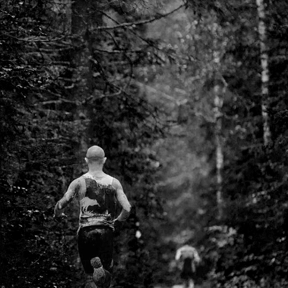
OFFIZIELLER PARTNER DES BRAVEHEART BATTLE 2019 Extreme Bedingungen – besondere Leistungen.
Die steilste Piste Thüringens, ein Halbmarathon über verschneite Berghänge und der Zieleinlauf durch eiskaltes Quellwasser: Wer das alles übersteht, erhält den verdienten Titel „Braveheart“.
Im März startet der Extrem-Geländelauf mit einem neuen Veranstalter und auf neuer Strecke durch. Ein Mix aus natürlichen und künstlichen Hindernissen auf dem teilweise unbefestigten Ski-Areal wird jedem Teilnehmer Höchstleistungen abverlangen – sowohl körperlich als auch mental.

WIR FREUEN UNS, ALS OFFIZIELLER PARTNERDABEI ZU SEIN: DANK UNSERER LANGJÄHRIGEN ERFAHRUNG IN DER BETREUUNG VON LEISTUNGSSPORTLERN WISSEN WIR GENAU, WORAUF ES WÄHREND EINES WETTKAMPFES UNTER EXTREMEN BEDINGUNGEN ANKOMMT.
Wir kennen die Herausforderungen und Risiken für Sportler bei einem solchen Ausnahme-Event und bieten den Teilnehmern des Braveheart Battle 2019 eine umfassende medizinische Betreuung durch die Ärzte des Curathleticum an.
Bei Verletzungen oder Beschwerden sind wir zur Stelle und übernehmen die Erstbehandlung vor Ort – so bleibt die Regenerationszeit möglichst gering und das Risiko von Folgeschäden sinkt.
ALLEN TEILNEHMERN WÜNSCHEN WIR VIEL ERFOLG UND SPASS BEIM BRAVEHEART BATTLE

SEPTEMBER 26, 2018
Conebeam
Röntgentechnik in drei Dimensionen Der SCS MedSeries® H22 bietet Bildgebung, wie sie sein sollte – mobil, schnell, zuverlässig.
Mit dem innovativen Scanner für digitale Volumentomographie bieten wir unseren Patienten im Curathleticum zahlreiche Vorteile. Ob schnelle Diagnose nach einer Verletzung, umfassende medizinische Analyse für Sportler oder Verlaufskontrolle nach einem chirurgischen Eingriff: Der SCS MedSeries H22 erlaubt ganz neue Wege der Diagnostik per 3D-Analyse.
Innerhalb weniger Sekunden fertigt der SCS MedSeries H22 Röntgenaufnahmen der Extremitäten an. Verletzungen an Kopf, Armen oder Beinen werden hochauflösend und dreidimensional sichtbar gemacht.
Als eine der ersten Praxen in Süddeutschland setzen wir diese Technologie ein, um vor Ort eine Diagnose zu stellen und den gesamte Behandlungsplan sofort entsprechend anzupassen.
Die Cone Beam-Volumentomographieerzeugt detaillierte 3D-Bilder mithilfe von kegelförmig ausgesandten Röntgenstrahlen. So werden zeitgleich zahlreiche Aufnahmen aus unterschiedlichen Winkeln angefertigt, aus denen anschließend ein dreidimensionales Modell berechnet wird.

Knochen und Gelenkstrukturen bildet der digitale Volumentomograph in hoher Auflösung ab: Knorpelschäden im Gelenk oder sehr feine Brüche können früh erkannt und dementsprechend therapiert werden. Die Strahlendosis einer Sitzung beträgt unter 50% der Strahlenmenge eines herkömmlichen Computertomographen.
Durch seine kompakte Bauweiseerlaubt der SCS MedSeries H22 die Untersuchung einzelner Extremitäten, während der Patient bequem sitzt. Das Liegen in einer beengten Röhre gehört dank der schwenkbaren und höhenverstellbaren Scannereinheit der Vergangenheit an.

Besonders interessant ist die neue Möglichkeitdes Belastungs-CT: Der Patient steht in der sogenannten Gantry, also der Scannereinheit, während die Röntgenbilder aufgezeichnet werden. Mit diesem Verfahren wird die Anatomie von Knie, Sprunggelenk oder Fuß unter natürlicher Belastung sichtbar gemacht und erlaubt neue Rückschlüsse oder gar Behandlungsansätze.
Als Zentrum für Sportmedizin und Unfallchirurgie setzen wir den SCS MedSeries H22 besonders nach Unfällen oder Verletzungen ein, um die betroffene Extremität schnell und umfassend zu beurteilen und eine zuverlässige Diagnose zu stellen.
Auch nach operativen Eingriffen nutzen wir die digitale Volumentomographie, um den Heilungsverlauf bei uns vor Ort zu kontrollieren und gegebenenfalls Ihren Therapieplan anzupassen.

Ihre Vorteile mit dem SCS MedSeries H22:
Kurze Untersuchungsdauer
Von der Konfiguration des Gerätes bis zur fertigen 3D-Aufnahme vergehen nur wenige Minuten.
Mobile Scannereinheit
Die Untersuchung erfolgt für Sie bequem, unkompliziert und ohne beengendes Gefühl. Wir haben den Scanvorgang jederzeit im Blick, sodass Fehler bei der Durchführung reduziert werden.
Niedrige Strahlendosis
Dank der neuartigen Technologie und kompakten Bauweise ist die Strahlungsbelastung um mindestens 50% niedriger als bei einer herkömmlichen Volumentomographie.
Schnelle Diagnose
Ihre Verletzung wird in kürzester Zeit erkannt und beurteilt. Den weiteren Behandlungsweg richten wir individuell auf Sie aus.
Untersuchung vor Ort
Wir führen die digitale Volumentomographie bei uns im Curathleticum durch – ohne Überweisung in die Radiologie: So entfallen für Sie Zeitaufwand, zusätzliche Wege und Wartezeiten.
Zuverlässige Ergebnisse
Gegenüber anderen Computertomographen erreicht der SCS MedSeries H22 eine mindestens 4x höhere Bildauflösung. Schon minimale Knorpelschäden oder feine Knochenbrüche können bei der ersten Untersuchung sicher erkannt werden.
Neuartige Untersuchungsmöglichkeiten
Dank der mobilen Scannereinheit sind Aufnahmen der unteren Extremität (Knie, Sprunggelenk & Knöchel, Fuß) auch unter Belastung möglich. So überprüfen wir z.B. nach einer Operation, ob die natürliche Funktion eines Gelenks wiederhergestellt ist.

SEPTEMBER 21, 2017
Hyaluronsäure: Einsatzmöglichkeiten
im Spitzensport
Gelenkschmerzen, verursacht durch degenerative Veränderungen, betreffen mit fortschreitendem Alter einen Großteil der Bevölkerung. Aber auch in jungen Jahren, speziell nach Verletzungen des Gelenks
Gelenkschmerzen, verursacht durch degenerative Veränderungen, betreffen mit fortschreitendem Alter einen Großteil der Bevölkerung. Aber auch in jungen Jahren, speziell nach Verletzungen des Gelenkknorpels, treten arthrotische Veränderungen auf, die zu Einschränkungen der Leistungsfähigkeit bei Spitzensportlern führen können.
Ein Artikel von PD Dr. Matthias Brem
Seit vielen Jahren werden unterschiedliche Hyalurosäurearten zur Therapie von degenerativen Gelenksveränderungen in der medizinischen Praxis verwendet {{1}}.
Hyaluronsäure (HA) ist ein natürlich vorkommendes Glykosaminoglykan, das aus einer Kette von Disacchariden (D-Glucuronsäure und N-Acetylglucosamin) besteht. Sie wird in hohen Konzentrationen in der Synovialflüssigkeit und im Bindegewebe gefunden {{1, 2}}. Im Gelenkknorpel übernimmt die HA eine entscheidende strukturgebende Rolle in der Knorpelmatrix.
Darüber hinaus hat sie wesentlichen Einfluss auf die Viscoelastizität der Synovialflüssigkeit {{1, 3}}. Im Rahmen der Arthrose scheinen komplexe Entzündungsprozesse eine das Fortschreiten negativ beeinflussende Rolle zu spielen {{4, 5}}. Einem Übersichtsartikel von Machado und Coautoren zufolge scheint die intraartikuläre Gabe von Hyaluronsäure Einfluss auf verschiedene Rezeptoren der Synovia auszuüben, die das Voranschreiten der Arthrose positiv beeinflussen können {{5}}. Edouart und Coautoren veröffentlichten einen Review über die Effektivität von HA-Injektionen nach traumatischen Verletzungen des Kniegelenks im Tiermodel.
In dem Übersichtsartikel konstatiert er, dass durch eine HA-Injektion nach Verletzungen des Meniskus und konsekutiver Meniskusteilresektion ein verbesserter Heilungsprozess und eine Schutzfunktion für den Knorpel erreicht werden kann. Laut Edouart waren nach vorderer Kreuzbandruptur die Effekte nicht mehr so ausgeprägt zu erkennen und die Effekte nach Knorpelschaden zumindest im Tierversuch zu vernachlässigen {{6}}.
Die intraartikuläre Gabe von Hyaluronsäure ist aus der täglichen medizinischen Praxis, speziell bei der Behandlung von Spitzenathleten, zum gegenwärtigen Zeitpunkt kaum außer Acht zu lassen. Persönliche Erfahrungen mit der Applikation von Hyaluronsäure nach Verletzungen des Knie- und Sprunggelenkes bestätigen die unterschiedlichen wissenschaftlichen Publikationen. In der Literatur sind mehrere Studien zu finden, die die Wirksamkeit der intraartikulären Gabe von Hyaluronsäure hinsichtlich der Schmerzreduktion und damit verbesserten Funktionalität des Gelenkes belegen {{1 – 3, 7 – 10}}.
Augenblicklich sind prinzipiell zwei unterschiedliche, pharmazeutisch aufbereitete HA-Typen erhältlich, die sich bezüglich des Molekulargewichts unterscheiden. (A) niedriges Molekulargewicht (0,5 – 3,6 million Da) und (B) hohes Molekulargewicht (≥ 6,0 million Da) {{4}}. Die pharmazeutische Aufbereitung scheint unterschiedlich wirksam hinsichtlich der biologischen Aktivität, der Verweildauer im Gelenk und der Schmerzreduktion zu sein {{7}}. Die Applikation im Gelenk stellt aber nur eine mögliche Anwendbarkeit der Hyaluronsäure dar. Bereits vor einem Jahrzehnt publizierte Petrella et al. eine Studie über den positiven Effekt der periartikulären Anwendung von Hyaluronsäure bei akuten Sprunggelenksdistorsionen {{11}}.
Weitere Einsatzmöglichkeiten
Ein weiterer Aspekt der Einsetzbarkeit der Hyaluronsäure, der zunehmend in den Fokus der Sportmedizin rückt ist die peritendinöse Injektion bei Tendopathien. Kaux et al. berichtet in einem Übersichtsartikel über unterschiedliche Studien, die sehr ermutigend sind, hinsichtlich des erfolgreichen Einsatzes der Hyaluronsäure bei Sehnenreizungen {{12}}.
Meine persönlichen Erfahrungen der letzten Jahre bezüglich der ultraschallgesteuerten Injektion bei Achillodynie, speziell bei Schnellkraft- und Ausdauersportlern, ist sehr positiv. Speziell die Kombination der Injektion von Hyaluronsäure mit fokussierter und radialer Stoßwellentherapie (ESWT) hat sich in meiner klinischen Anwendung bewährt.
Experimentelle Untersuchungen konnten zeigen, dass durch die ESWT eine vermehrte Expression von Lubricin hervorgerufen wird {{13}}. Die damit verbundene Verbesserung der Gleitfähigkeit des Sehnengewebes scheint durch die Kombination mit Hyaluronsäre nochmals verbessert zu werden. Wobei die endgültige Wirkungsweise im Bezug auf das Zusammenspiel von ESWT und Hyaluronsärenjektion sicherlich noch weiterer Untersuchungen bedarf. Die bei Achillodynie vorhanden Schmerzen jedoch können auch bei Topathleten unter Vermeidung eines wesentlichen Trainingsausfalles reduziert werden und die Leistungsfähigkeit erhalten werden.
Die Literaturliste können Sie unter info@thesportgroup.de anfordern.
SEPTEMBER 21, 2017
Knorpel-
verletzungen
bei Leistungs-
sportlern
Welche operativen Therapien und Optionen in der Nachbehandlung den „Return to Sports“ beschleunigen
Ein Artikel von PD Dr. med. Matthias Brem, Dr. med. Hermann Josef Bail und Dr. med. Johannes Pauser
Die Behandlung eines frischen traumatischen Knorpelschadens ist für das medizinische Team in allen Belangen eine große Herausforderung. Die operativen Behandlungsmethoden sind in vielen klinischen und experimentellen Studien untersucht worden. Die klinische Evidenz ist ein entscheidendes Kriterium für die Wahl des Verfahrens. Die postoperative Ausfallzeit während der Rehabilitation ist für viele professionelle Sportler ein ebenso gewichtiges Argument. Eine ausführliche Aufklärung über unterschiedliche Behandlungsmethoden sollte die Entscheidung beeinflussen, welches Verfahren gewählt wird. Sie muss individuell auf die Bedürfnisse und Wünsche des Patienten zugeschnitten sein.
In der Literatur sind für unterschiedliche Verfahren zahlreiche Studien vorhanden. Empfohlen wird derzeit die operative Intervention, da der verletze Knorpel nur eine limitierte Selbstheilungstendenz hat und das Risiko einer frühzeitigen Arthroseentwicklung besteht.
Das wohl am häufigsten angewandte reparative Verfahren ist die Mikrofrakturierung. In der von John Richard Steadman (2001) beschriebenen Technik wird nach einem sorgfältigen Debridement des Defekts, der Abtragung der Sklerosezone am Defektgrund und der Schaffung von stabilen Knorpelrändern der subchondrale Knochen mit gebogenen Ahlen perforiert. Dabei sollen Knochenmarkzellen freigesetzt werden, die anschließend zu einem faserknorpeligen Gewebe ausdifferenzieren.
Dieses Verfahren zeichnet sich durch seine schnelle Durchführbarkeit, die kostengünstige Handhabung und die relativ schnelle Rückkehr des Athleten zu seiner sportlichen Belastbarkeit aus. A.B. Campbell beschreibt in seinem Übersichtsartikel (2015) eine Wahrscheinlichkeit von 75 Prozent, dass ein Profisportler in den Sport zurückkehrt. Die Quote der Sportler, die das sportliche Level wieder erreichen, wird von ihm mit ca. 69 Prozent angeben. Die kurzfristigen Ergebnisse sind sicherlich zufriedenstellend, im langfristigen Follow-up jedoch verschlechtert sich die Schmerz- und Aktivitätssituation der Patienten wieder. Als mögliche Ursachen können eine mindere Qualität des Faserknorpels, die hohe Belastung durch Scherkräfte und die sehr schnelle Rückkehr zur Belastung im Sport angenommen werden.
Knorpel-Knochen-Zylinder
Eine weitere operative Technik ist die OAT (Ostechondral Autograft), bei der an einer außerhalb der Belastungszone gelegenen Stelle des Gelenks Knorpel-Knochen-Zylinder entnommen und in den Defekt transplantiert werden. Dies kann als einzelnes Transplantat oder durch mehrere Transplantate als Mosaikplastik erfolgen. Die „Return to Sports“-Wahrscheinlichkeit liegt laut Literaturangaben bei 89 Prozent, jedoch erreichen nur ca. 70 Prozent der Hochleistungssportler das gleiche Level an sportlicher Belastbarkeit wie vor dem Unfall.
In den vergangenen Jahren wurden zunehmend Studien und Ergebnisse der Autologen Chondrozyten Transplantation (ACT oder engl. ACI) veröffentlicht, die Hoffnung machen, gute mittel- bis langfristige Ergebnisse zu erzielen. Bei dieser Technik werden Chondrozyten aus dem Gelenk entnommen und in vitro angezüchtet und vermehrt. In einer zweiten Operation werden die Zellen in den Knorpeldefekt wieder implantiert. Die Chondrozytenlösung wird entweder unter eine Kollagenmembran oder in früher beschriebenen Verfahren unter einen Periostlappen, der in den Defekt eingenäht wird, eingebracht.
Eine Weiterentwicklung dieser ursprünglich beschriebenen Technik ist das Matrix-assoziierte Transplantationsverfahren (MACT), bei dem die im Labor vervielfältigten Zellen auf eine Kollagen-Trägersubstanz aufgebracht werden. Unterschiedliche Hersteller bieten diese Form der Chondrozytentransplantation an. Klinische Ergebnisse zeigen ermutigende Resultate hinsichtlich der Erfolgsrate dieses Verfahrens. Im bereits zitierten Review von Campbell wird die „Return zu Sports“-Rate der Spitzensportler mit 84 Prozent angegeben. Das Vorverletzungslevel erreichten immerhin 76 Prozent aller Sportler, die in diese Untersuchung eingeschlossen wurden.
Kollagenmatrix als Vlies
Eine andere operative Herangehensweise an den Knorpeldefekt ist die Verwendung der Trägersubstanzen, zum Beispiel Kollagenvliese ohne zelluläre Anreicherung. Eine Variante stellt eine modifizierte Mikrofrakturierung dar, bei der nach der Mikrofrakturierung eine Kollagenmatrix als Vlies oder als flüssige Kollagensubstanz, wie beispielsweise eine selbstaushärtende Suspension aus Atellocollagen und Fibrinogen, in den Defekt eingebracht wird (z.B. CartiFillTM der Firma RMS).
Damit wird das Blut-Zell-Gemisch an der Defektstelle fixiert und soll so zu einer Ausdifferenzierung von Knorpelregeneratgewebe führen. Bei diesem Verfahren wird nach der Mikrofrakturierung die Spülflüssigkeit aus dem Kniegelenk entfernt, der Defektgrund getrocknet und die Suspension mittels Spritze in den Defekt eingebracht. Die Suspension verfestigt sich nach wenigen Minuten und ist stabil in den Knorpeldefekt eingebracht. Die ersten publizierten Ergebnisse scheinen sehr erfolgsversprechend zu sein (signifikante Verbesserung des Lysholm scores und MOCART scores im Zwei-Jahres-Follow-up). In einer Bildgebungsstudie konnten im Zwei-Jahres-Follow-up ebenfalls erfolgversprechende Ergebnisse gezeigt werden. Der Vorteil dieser Methode ist die einzeitige Operation.
Schnellster „Return to Sports“
In der Literatur werden für die einzelnen genannten Operationsverfahren unterschiedliche Zeiten angegeben, wann ein Leistungssportler wieder seine gewohnte Belastung im Sinne des „Return to Sports“ aufnehmen kann. In einer vergleichenden Zusammenfassung scheint die OAT die schnellste Wiederaufnahme des Sports nach durchschnittlich etwa 7,1 Monaten zu ermöglichen. Die Mikrofrakturierung ermöglicht dem betroffenen Athleten nach ca. 8,6 Monaten und die ACT nach ca. 16 Monaten eine Wiederaufnahme des Sports.
Bei allen in publizierten Studien verfügbaren Ergebnissen sind immer die Defektlokalisation und die Größe des Defekts sowie Faktoren wie Beinachse und Begleitverletzungen in die Überlegungen mit einzubeziehen. Daher sind die in der Literatur verfügbaren Ergebnisse sehr heterogen und somit oftmals schwer zu vergleichen.
Nachbehandlungsschema
Das postoperative Vorgehen und die Weiterbehandlung sind in der Literatur ebenso heterogen beschrieben. Ein Nachbehandlungsschema für Patienten, die mittels MACT behandelt wurden, ist von der Arbeitsgruppe „Klinische Geweberegeneration“ der Deutschen Gesellschaft für Unfallchirurgie (DGU) und der Deutschen Gesellschaft für Orthopädie und orthopädische Chirurgie (DOOC) verfasst und im Jahr 2014 veröffentlicht worden. In der Zusammenfassung der Empfehlung wird darauf verwiesen, dass klare Nachbehandlungsschemata nach MACT am Kniegelenk nicht existieren und weiterer Bedarf an Optimierung sowie an Datenerfassung bestehe.
Ein Konsensus besteht aber im Hinblick auf den Belastungsaufbau nach femoralen Knorpelschäden, die die Belastung auf Bodenkontakt für sechs Wochen limitiert und eine passive Bewegung mittels CPM für 3-8 h/Tag nach Redonzug empfiehlt. Im Anschluss sollte ein sukzessiver Belastungsaufbau erfolgen. Eine Bewegungslimitierung ist bei femoralen Defekten nicht notwendig. Bei retropatellaren Defekten wird jedoch eine Bewegungslimitierung in Woche 1 bis 2: 0-30°, Woche 3 bis 4: 0-60° und Woche 5 bis 6: 0-80°empfohlen, wobei eine Vollbelastung in Streckstellung möglich ist.
In der Nachbehandlung von Knorpelschäden sind unterschiedliche Varianten in der Diskussion. Eine von Mustafa Karakaplan veröffentlichte Studie (2015) konnte im Tierversuch einen positiven Effekt auf die Knorpelregeneratbildung unter der Gabe von ACP und Mikrofrakturierung im Vergleich zu alleiniger Mikrofrakturierung zeigen. Weitere Untersuchungen im klinischen Setting bei unterschiedlichen Operationstechniken sollten durchgeführt werden, um diesen positiven Einfluss auch beim Menschen zu evaluieren.
Ein anderer Ansatz kann die postoperative Gabe von Hyaluronsäure zur Verbesserung der Gelenkshomöostase und zur Viscosupplementation des Gelenks sowie zur möglichen Entzündungsreduktion sein. Die Weiterentwicklung verschiedener Hyaluronsäureprodukte, die die Eigenschaften von unterschiedlich aufgearbeiteten Präparaten verbinden, kann möglicherweise einen positiven Einfluss auf das Outcome nach knorpelchirurgischer Behandlung haben. Wissenschaftliche Daten stehen hierzu jedoch noch aus.
Fallbericht
Ein Beispiel aus unserer Klinik: Eine 22 Jahre alte Fußballspielerin erlitt eine Ruptur des Vorderen Kreuzbands (VKB) und einen viergradigen, bereits durch Mikrofraktur vorbehandelten femoralen Knorpelschaden. In der Vorgeschichte wurde die Patientin bereits nach einer Meniskusläsion und einem traumatischen Knorpelschaden operativ mittels einer Meniskusteilresektion und einer Mikrofrakturierung in Südamerika behandelt.
Die Patientin wurde in einer operativen Sitzung behandelt. Eine Rekonstruktion des VKB erfolgte mittels vierfach gefaltetem Sehnentransplantat M. semitendinosus, einer Mikrofrakturierung und Auffüllung des Defekts mit CartiFill. In der Nachbehandlung fand eine Teilbelastung mit Abstellen des Beins für sechs Wochen und eine CPM-Mobilisierung für 6h/Tag bis 90° Beugung statt. Im kurzzeitigen Nachuntersuchungsintervall ist die Patientin vollkommen beschwerdefrei.
PD Dr. med. Matthias Brem ist Oberarzt der Klinik für Orthopädie und Unfallchirurgie am Klinikum Nürnberg. Der Facharzt betreut mehrere Teams im Leistungssport, u.a. den Fußball-Zweitligisten 1. FC Nürnberg, die Basketball-Bundesligamannschaft rent4office Nürnberg und die Bundesligamannschaft der Ringer „Johannis Grizzlys“
Professor Dr. med. Hermann Josef Bail leitet die Universitätsklinik für Orthopädie und Unfallchirurgie am Klinikum Nürnberg. Er ist Facharzt für Chirurgie mit dem Schwerpunkt Unfallchirurgie.
Dr. med. Johannes Pauser, Oberarzt der Chirurgischen Notaufnahme am Klinikum Nürnberg, ist Facharzt für Orthopädie und Unfallchirurgie mit zusätzlicher Qualifikation in u.a. Chirotherapie, Notfallmedizin und Sportmedizin. Er ist Mannschaftsarzt der Basketball-Bundesligamannschaft rent4office Nürnberg.
Quelle: sportaerztezeitung.de
Eine Literaturliste zu diesem Artikel finden Sie hier.
SEPTEMBER 18, 2017
Was wissen wir
über Triggerpunkte?
Die Wissenschaft streitet, belegbare Kenntnisse gibt es kaum.Autor Prof. Dr. Christoph Schmitz fordert einen neuen Ansatz in der Forschung:
Die Behandlung von Triggerpunkten (oder etwas allgemeiner: des myofaszialen Schmerzsyndroms) ist aus der modernen Sportmedizin nicht mehr wegzudenken. Eine Suche bei Google ergab für die Stichworte „Triggerpunkte Sportmedizin“ mehr als 27.000 Ergebnisse und für die Stichworte „myofaszial Sportmedizin“ über 9000 Ergebnisse (Stand 31.12.15).
Da sollte es doch eigentlich nicht schwerfallen, die wesentlichen Erkenntnisse zur Ätiologie (den auslösenden Ursachen), Pathophysiologie (den Veränderungen im Gewebe), Diagnostik und Therapie von Triggerpunkten bzw. zum myofaszialen Schmerzsyndrom (im folgenden TrP/MFS abgekürzt) schnell zusammen zu haben.
Tatsächlich sieht die Realität aber völlig anders aus. Beginnen wir unsere Recherche in einem Medium, das heutzutage von sehr vielen Menschen (d.h. Patientinnen und Patienten) zuerst als Lexikon bzw. Lexikonersatz herangezogen wird: Wikipedia. Interessanterweise gibt es auf der deutschsprachigen Seite von Wikipedia keinen Eintrag zu „Triggerpunkten“, wohl aber zu „Triggerpunkttherapie“. Wie weiter unten ausgeführt wird, könnte dieser Umstand die gegenwärtige Situation nicht besser widerspiegeln: Wir behandeln zwar etwas, wissen aber nicht genau, was das tatsächlich ist. Viel ist bei Wikipedia auch nicht zu erfahren. Triggerpunkte „sind lokal begrenzte Muskelverhärtungen in der Skelettmuskulatur, die lokal druckempfindlich sind und von denen übertragene Schmerzen ausgehen können“. Dies ist nicht nur wenig, sondern schlicht zu wenig.
Es gibt eine viel bessere Definition aus einer kürzlich publizierten Arbeit von Quintner et al. (2015), auf die wir weiter unten noch detailliert zu sprechen kommen: „The theory of MPS comprised two essential components: the TrP, a localized area of tenderness or hyperirritability deep within voluntary muscle; and a predictable discrete zone of deep aching pain, which could be located in the immediate region of or remote from the TrP, and which was worsened by palpation of the TrP.“
Anatomischer Zusammenhang
Wichtig erscheint hier die Formulierung „predictable discrete zone of deep aching pain“. Mit anderen Worten, die übertragenen Schmerzen treten nicht einfach irgendwo auf, sondern in einer voraussagbaren und damit reproduzierbaren Körperregion, und sie können durch Druck auf den zugehörigen Triggerpunkt gesteigert werden.
Dies bedeutet jedoch, dass es irgendeine Form von anatomischem Zusammenhang zwischen dem Triggerpunkt selbst und der Zone des übertragenen Schmerzes geben muss. Dabei muss keineswegs ein direkter anatomischer Zusammenhang bestehen, zum Beispiel ein Nerv, der einen Muskel durchquert und durch Kontraktion des Muskels in Mitleidenschaft gezogen wird (was tatsächlich für die allermeisten Triggerpunkte auch nicht bekannt ist). Vielmehr könnte der übertragene Schmerz einem ähnlichen Konzept wie dem der Head‘schen Zonen folgen, die ja zum Beispiel für den Herzinfarkt (Schmerzen an der Innenseite des linken Arms) gut bekannt sind. Allerdings basiert das Konzept der Head’schen Zonen auf Querverbindungen zwischen dem somatischen und dem vegetativen Nervensystem, was für den übertragenen Schmerz bei TrP/MFS nicht zutrifft (hier liegen sowohl die Triggerpunkte als auch die Regionen des übertragenen Schmerzes im Bereich des somatischen Nervensystems).
Bevor wir uns im Detail mit dem Phänomen TrP/MFS gerade in der modernen Sportmedizin beschäftigen, werfen wir zunächst einen kurzen Blick auf die Häufigkeit dieser Problematik. Bei Wikipedia heißt es: „Rund 80 bis 90 Prozent der Schmerzsyndrome sollen auf derartige Muskulaturverhärtungen zurückzuführen sein.“ Wikipedia gibt keine Quelle für diese Zahlen an. Sie passen allerdings gut zu seriösen Zahlen von Murray et al. (2013), die im Journal of the American Medical Association (JAMA) ein sogenanntes Disability-Adjusted Life-Year (DALY) Ranking, also eine Art negative „Hitparade“, der Top-30-Erkrankungen und Verletzungen in den USA zusammengestellt haben.
Bis heute ist es nicht gelungen, die Ätiologie und Pathogenese von Triggerpunkten bzw. dem myofaszialen Schmerzsyndrom aufzuklären. Weder bildgebende Verfahren, noch biochemische Analysen, Biopsien oder Post-mortem-Untersuchungen konnten dazu beitragen. Darum brauchen wir dringend einen neuen, ganzheitlich- wissenschaftlichen Ansatz zur Beschäftigung mit dem Phänomen.
Das DALY Ranking
Für das Jahr 2010 sah das DALY Ranking für die USA auf den ersten 12 Plätzen wie folgt aus:
1: Ischämische Herzerkrankungen
2: Chronisch obstrukive Lungenerkrankungen
3: Lendenwirbelschmerzen
4: Lungenkrebs
5: Depressionen
6: Andere Erkrankungen des Stütz- und Bewegungsapparates
7: Schlaganfall
8: Diabetes
9: Verkehrsunfälle
10: Drogenmissbrauch
11: Nackenschmerzen
12: Alzheimer-Erkrankung
Da sowohl bei Tendinopathien als auch bei Lendenwirbelschmerzen und Nackenschmerzen Triggerpunkte eine wesentliche Rolle spielen sollen (Google-Suche am 31.12.2015 nach „Rückenschmerzen Triggerpunkte“: knapp 18.000 Ergebnisse), kann man davon ausgehen, dass es TrP/MFS im DALY Ranking für die USA in die Top 10 schaffen würden – vorausgesetzt, es gäbe umfangreiches Wissen zu Ätiologie und Pathogenese von TrP/MFS.
Genau dies ist aber nicht der Fall. Vielmehr würde nach streng wissenschaftlichen Kriterien das gegenwärtige Urteil über die Ätiologie und Pathogenese von TrP/MFS sehr kurz ausfallen: Beides ist letztlich unbekannt! In diesem Zusammenhang sei auf die bereits oben erwähnte Arbeit von Quintner et al. verwiesen, die alle gelesen haben sollten, die irgendetwas mit TrP/MFS zu tun haben (frei verfügbar unter www.painaustralia. org.au/images/pain_australia/Rheumatology- 2014-Quintner-rheumatology_keu471. pdf)*1. Quintner et al. kommen zu einem vernichtenden Ergebnis: „We find that both {{d.h., TrP und MFS}} are inventions that have no scientific basis, whether from experimental approaches that interrogate the suspect tissue or empirical approaches that assess the outcome of treatments predicated on presumed pathology. Therefore, the theory of MPS {{myofascial pain syndome, also MFS}} caused by TrPs has been refuted.“ Natürlich ist dies eine sehr provokativ formulierte Einschätzung, und die Reaktion ließ nicht lange auf sich warten.
Schon kurze Zeit später publizierten Dommerholt und Gerwin (2015) einen detaillierten Kommentar zu der Arbeit von Quintner et al., in der sie zu einem nicht minder vernichtenden Urteil kamen: „The current paper demonstrates that the Quintner et al. paper is a biased review of the literature replete with unsupported opinions and accusations. In summary, Quintner et al. have not presented any convincing evidence to believe that the Integrated TrP Hypothesis should be laid to rest.“ Auch diesen Kommentar von Dommerholt und Gerwin sollten alle gelesen haben, die sich mit TrP/MFS beschäftigen (frei verfügbar online)*2.
Streit in der Literatur
Bei diesem heftigen Streit in der wissenschaftlichen Literatur geht es um genau das, was eingangs bereits ausgeführt wurde: Wir behandeln TrP/MFS, wissen aber nicht genau, was TrP/MFS tatsächlich sind.
Es ist bis heute nicht gelungen, die Ätiologie und Pathogenese von TrP/MFS mit bildgebenden Verfahren, biochemischen Analysen, Biopsien oder Post-mortem- Untersuchungen aufzuklären (Details finden sich bei Quintner et al. und Dommerholt und Gerwin). Dies gilt auch für die neuerdings stark propagierte Ultraschall-Elastografie (siehe z.B. Turo et al., 2015). Grob gesagt wird bei diesem Verfahren ein Gewebe (inneres Organ, Muskel) in unterschiedlichen Spannungs- oder Dehnungszuständen geschallt und Veränderungen im Vergleich zu anderem Gewebe (z.B. der gesunden Gegenseite) oder im Vergleich vor und nach einer Behandlung analysiert. Es ist völlig richtig, dass man mit Ultraschall- Elastografie veränderte Muskelspannung nach einer Behandlung nachweisen kann – aber dies besagt eben nicht, dass dieses Verfahren zum bildgebenden Nachweis von TrP/ MFS geeignet ist.
Insgesamt gibt es bis heute Parameter biologischer Prozesse, die prognostische oder diagnostische Aussagekraft für das Vorliegen oder Nicht-Vorhandensein von TrP/ MFS haben.
Tiermodelle funktionieren nicht
Darüber hinaus gibt es kein einziges auch nur halbwegs brauchbares Tiermodell zu TrP/ MFS, was in dreifacher Hinsicht zum Nachdenken anregen sollte: (i) Das Fehlen geeigneter Tiermodelle ist einer der Hauptgründe dafür, dass sich die Grundlagenforschung so wenig mit TrP/MFS beschäftigt – viel zu wenig, wenn man die oben genannten Zahlen von Murray et al. zum DALY Ranking bedenkt. (ii) Ein vollständiges Tiermodell zu TrP/MFS kann und wird es aber niemals geben, denn das Phänomen von übertragenen Schmerzen, die durch Druck auf den zugehörigen Triggerpunkt gesteigert werden, kann im Tiermodell nicht nachgestellt werden. (iii)
Aller Wahrscheinlichkeit nach entwickeln Tiere keine TrP/MFS, da sie nicht im Büro sitzen, aber auch keinen Sport treiben. Der letzte Aspekt mag ungewöhnlich erscheinen, ist aber von ganz entscheidender Bedeutung für die Auseinandersetzung mit TrP/MFS in der modernen Sportmedizin. Konkret wird hier die Frage nach der „physiologischen“ Verwendung des Stütz- und Bewegungsapparates beim Menschen angesprochen, d.h. der Primärprävention von TrP/MFS.
Die populärwissenschaftliche Literatur ist in dieser Hinsicht voll von Ratschlägen (die Beschreibung „gute Ratschläge“ wird hier bewusst vermieden), die aber allesamt einen ganz wichtigen Aspekt vergessen: Aller Wahrscheinlichkeit nach macht die in der Evolution einzigartige kognitive Entwicklung des Menschen eine „physiologische“ Verwendung seines Stütz- und Bewegungsapparates gar nicht mehr möglich.
„Abreagieren“ ist unnatürlich
Um nur ein Beispiel zu geben: Das „Abreagieren“ zum Beispiel bei einer intensiven Partie Squash (mit all ihren möglichen negativen sportmedizinischen Konsequenzen) nach stundenlangen zermürbenden Verhandlungen im Büro wird von vielen Menschen als wohltuend empfunden, ist aber in der Natur auch nur annähernd ohne Beispiel. Von daher greift die Idee, sich in der Grundlagenforschung wegen des Fehlens von Tiermodellen nicht oder nur kaum mit TrP/MFS zu beschäftigen, viel zu kurz.
Was wir tatsächlich dringend brauchen, ist ein neuer, ganzheitlich-wissenschaftlicher Ansatz zur Beschäftigung mit dem Phänomen TrP/MFS, der weit über das hinausgeht, was Quintner et al. an Evidenz herangezogen haben, um das Konzept TrP/MFS in ihren Augen zu widerlegen – der allerdings auch weit über das hinausgehen muss, was Dommerholt und Gerwin in ihrer Reaktion auf Quintner et al. zusammengetragen haben, um das Konzept TrP/MFS zu verteidigen.
Wie ein solcher neuer, ganzheitlich-wissenschaftlicher Ansatz aussehen könnte, wird an anderer Stelle diskutiert werden. Es sei hier jedoch auf die Erfolgsgeschichten der Psychoneuroendokrinologie (derzeit über 4000 Einträge in „PubMed“) sowie der Psychoneuroimmunologie (derzeit über 2000 Einträge in „PubMed“) verwiesen, die auf ähnlichen neuen, ganzheitlich-wissenschaftlichen Ansätzen basieren.
Zwei wichtige Fragen
Zusammenfassend ergeben sich aus dem oben beschriebenen Streit zwischen Quintner et al. und Dommerholt und Gerwin zum jetzigen Zeitpunkt insbesondere die beiden folgenden Fragen:
• Wie kann man Kriterien für valide und reproduzierbare Diagnostik von TrP/MFS erstellen, wenn man eigentlich gar nicht weiß, was da genau diagnostiziert werden soll?
Quintner et al. zitieren in diesem Zusammenhang eine Arbeit von Tough et al. (2007), nach denen in der Literatur mindestens 19 verschiedene Kriterien zur Diagnose von TrP/ MFS beschrieben wurden. Darüber hinaus verweisen sie auf eine Reihe von Studien, in denen verschiedene Untersucher bei denselben Patienten zu unterschiedlichen Diagnosen kamen. Insgesamt muss die Schlussfolgerung von Quintner et al. – „physical examination cannot be relied upon to diagnose a condition that is supposed to be defined by that physical examination. That is, the pathognomonic criterion for making the diagnosis of MPS is unreliable“ – sehr ernst genommen werden.
Sie darf aber nicht zum Verwerfen des Konzepts TrP/MFS führen, sondern sollte als Herausforderung für den oben beschriebenen neuen, ganzheitlich-wissenschaftlichen Ansatz zur Beschäftigung mit dem Phänomen TrP/MFS verstanden werden. Alle, die heute TrP/MFS diagnostizieren und behandeln, sollten sich der Kritik von Quintner et al. bewusst sein, denn früher oder später wird diese Kritik bei den Patientinnen und Patienten ankommen und dann evtl. eine Rechtfertigungsdebatte auslösen, die in den USA evtl. schon begonnen hat und die heftige Gegenreaktion von Dommerholt und Gerwin erklären könnte (bei der übrigens auch handfeste kommerzielle Gründe eine Rolle gespielt haben können; siehe das Statement of Interests bei Dommerholt und Gerwin).
• Nach welchen Kriterien werden gerade in der modernen Sportmedizin Therapieoptionen für TrP/MFS bewertet und ausgewählt, wenn man eigentlich gar nicht weiß, was da genau behandelt wird?
Diese Frage ist fast noch kritischer als die Frage nach der reproduzierbaren Diagnostik von TrP/MFS. Man sollte sich hüten, die Diskussion an dieser Stelle mit dem Verweis auf „Wer heilt, hat Recht!“ abzubrechen. Fragt man nämlich zurück, „Wie wird denn Heilung in diesem Fall definiert?“ und bekommt dann „mittels Schmerzfreiheit“ als Antwort zurück, liegt man gerade in der Sportmedizin komplett falsch. Kreutz (2015) hat dies kürzlich in einer hervorragenden Arbeit auf den Punkt gebracht: „Die Behandlung von Triggerpunkten mit dem alleinigen Ziel der Schmerzfreiheit ist insbesondere im Spitzensport unzureichend. Schmerz stellt weder ein Frühwarnsystem noch einen suffizienten Verlaufsparameter auf dem Weg zur Systemnormalisierung dar. Auch latente (im Alltag nicht spürbare) mTP {{myofasziale Triggerpunkte}} stören die für die sportliche Leistungsfähigkeit wesentlichen Parameter wie Propriozeption und Muskelaktivitätsmuster. Zudem führen sie zur Muskelschwäche ohne Atrophie nicht nur des betroffenen Muskels, sondern ganzer funktioneller Ketten.“ (frei verfügbar unter www.medicalsportsnetwork.com/medical/2564475818/Dr-Andreas-Kreutz/Myofasziale-Triggerpunkte.html)*3.
Fazit
Kurz gesagt: Wir brauchen eine neue, intensive Auseinandersetzung mit dem Phänomen TrP/MFS in der modernen Sportmedizin, einschließlich einem neuen, ganzheitlich-wissenschaftlichen Ansatz, vergleichbar zum Beispiel mit dem der Psychoneuroendokrinologie. Schon heute stehen verschiedene Therapieverfahren zur Verfügung, um in dem von Kreutz formulierten Konzept gemeinsam zur Anwendung zu kommen. Diese werden in der nächsten Ausgabe der sportärztezeitung vorgestellt und kritisch gegeneinander abgewogen.
Univ. Prof. Dr.med. Christoph Schmitz leitet den Lehrstuhl für Anatomie II an der Ludwig-Maximilians- Universität München.
Eines seiner Forschungsgebiete befasst sich mit innovativen Akut-Behandlungen bei Verletzungen von Profi-Fußballspielern.
Quelle: sportaerztezeitung 01/2016
Eine Literaturliste erhalten Sie beim Verfasser.
SEPTEMBER 18, 2017
Brennpunkt Muskel: Verletzungen im Sport und Relevanz außerhalb des Leistungssports
Muskelverletzungen gehören zu den häufigsten Schäden im Leistungs- und Breitensport.
Im Profifußball entfallen z. B. rund ein Drittel aller Verletzungen auf Muskelverletzungen.
Einer aktuellen Untersuchung nach sind die häufigsten Verletzungen in der 1. Fußball-Bundesliga bis zur Winterpause 2016/17 mit > 2 Wochen Pause Muskelfaserriss Oberschenkel (15x) gefolgt von Achillessehnenproblemen, Adduktorenzerrungen, Leistenproblemen und Muskelfaserrissen im Adduktorenbereich (jeweils 7x) – siehe www.fussballverletzungen.com.
Ein Interview mit Univ.-Prof. Dr. med. Christoph Schmitz
Zu weiteren „Risikosportarten“ zählen u.a. Handball (siehe VBG-Sportreport 2016), aber auch Tennis, Leichtathletik, Volleyball usw. Damit aber nicht genug. Laut einem Bericht der DAK-Gesundheit sind Rückenleiden und andere Muskel-Skelett-Erkrankungen der häufigste Grund für Krankmeldungen deutscher Arbeitnehmer (22 %). Im November 2016 haben wir von der sportärztezeitung auf unserem Symposium Fußballmedizin & Konservative Sportmedizin den Bereich Muskel als einen unserer Hauptthemen gewählt. Dort ist auch die Idee entstanden, den Fokus einer Ausgabe der sportärztezeitung auf das Thema Muskel zu legen. Da Prävention, Regeneration und konservative Therapieoptionen in diesem Bereich eine ganz entscheidende Rolle spielen, stellen wir Ihnen in dieser Ausgabe unterschiedliche Aspekte vor. Den Anfang macht ein Interview, das Robert Erbeldinger mit Univ.-Prof. Dr. med. Christoph Schmitz, Inhaber des Lehrstuhls für Anatomie II an der Ludwig-Maximilians- Universität München und Wissenschaftlicher Beirat der sportärztezeitung, über Aspekte von Muskelverletzungen im Sport führte. Der Fokus lag dabei auf besseren Therapiemöglichkeiten durch verfeinerte Einteilung und relevantere Tiermodelle.
Lieber Christoph, Du hast bei unserem Symposium „Fußballmedizin & Konservative Sportmedizin“ in der ARCUS Sportklinik Pforzheim im November 2016 einen bemerkenswerten Vortrag zum Thema „Anatomie einer Muskelverletzung” gehalten, bei dem Du unter anderem eine Verfeinerung der Einteilung von Muskelverletzungen nach Dr. Müller-Wohlfahrt und Kollegen vorgeschlagen hast. Um was geht es genau?
Kurz gesagt erscheint mir die Einteilung der teilweisen Muskelrisse in Typ 3a (≤ 5 mm; intrafaszikulär) und Typ 3b (≥ 5 mm; interfaszikulär) zu ungenau. Aus anatomischer Sicht sehe ich zunächst keine Begründung für eine Grenze bei 5 mm. Ich konnte auch keine Literatur finden, welche die Grenze bei 5 mm durch Biopsien validiert. Darüber hinaus ist mir unklar, wie diese Grenze von 5 mm in der Klinik erkannt werden soll.
Ist das nur eine akademische Frage oder ist diese Diskussion tatsächlich von klinischer Relevanz?
Eindeutig letzteres. Dazu ein aktuelles Beispiel. Kollegen aus den Niederlanden (die sogenannte „Dutch Hamstring Injection Therapy (HIT) Study Investigators“) haben in 2014 im New England Journal of Medicine eine Studie zur Therapie von akuten Verletzungen der ischiokruralen Muskulatur mit platelet rich plasma (PRP) publiziert {{1}}. Die Zeit zur Wiederaufnahme sportlicher Aktivitäten schwankte zwischen ca. 10 und 180 Tagen, wobei kein statistisch signifikanter Unterschied zwischen PRP und Kochsalzinjektion gefunden wurde. Nimmt man aber die Kriterien von Dr. Müller-Wohlfahrt und Kollegen (Typ 3a: 10 – 14 Tage bei optimaler Behandlung; Typ 3b: ungefähr sechs Wochen bei optimaler Behandlung), waren das in der niederländischen Studie praktisch ausnahmslos Typ 3b Verletzungen. Aus wissenschaftlicher Sicht macht das wenig Sinn. Wenn die Zeit zur Wiederaufnahme sportlicher Aktivitäten derart unterschiedlich ist, sollte man eher daran denken, dass hier nicht die gleichen Verletzungen vorgelegen haben. Dies hätte auch wichtige Konsequenzen für mögliche Therapieoptionen.
Damit kommen wir zum zweiten Teil Deines Vortrags. Neue Therapieoptionen werden ja oftmals zunächst im Tiermodell getestet. Gibt es da etwas Neues?
Ja, aber da war ich schon perplex. Kollegen aus Salzburg haben kürzlich im American Journal of Sports Medicine eine experimentelle Studie zur Beschleunigung von Muskelregeneration mit extrakorporalen Stoßwellen publiziert {{2}}.
Dein Spezialgebiet.
Ja. Aber als Modell für Muskelschäden wurde in dieser Studie die Injektion eines bestimmten Kardiotoxins (CTX) in den M. quadriceps femoris von Ratten verwendet – also eine chemisch induzierte Muskelverletzung.
Wo liegt das Problem?
Man weiß aus der Literatur {{3}}, dass sich CTX-induzierte Muskelschäden von mechanisch induzierten Muskelschäden in der Dynamik der Muskelregeneration unterscheiden. Oder anders gesagt: CTX-induzierte Muskelschäden sind für die Entwicklung innovativer Therapiemöglichkeiten für Muskelverletzungen im Sport eventuell nur von eingeschränkter Relevanz.
Gibt es denn relevantere Tiermodelle?
Durchaus. Man müsste nur die gute alte, experimentell induzierte „controlled strain injury“ wieder im Labor etablieren. Wesentliche Arbeiten {{4, 5}} zu dieser Methode wurden übrigens vor ca. 20 Jahren von einer Arbeitsgruppe an der Duke University in Durham (North Carolina, USA) um Dr. William E. Garrett Jr. publiziert, der bei der englischen Ausgabe des Buchs von Dr. Müller-Wohlfahrt und Kollegen zum Thema „Muskelverletzungen im Sport“ als Mitautor und Co-Herausgeber mitgewirkt hat.
Gibt es Hinweise darauf, dass die experimentell induzierte „controlled strain injury“ tatsächlich relevanter ist als z. B. CTX-induzierte Muskelschäden?
Absolut. Für die experimentell induzierte „controlled strain injury“ konnte z. B. gezeigt werden, dass der entstehende Muskelschaden keine lineare Funktion der einwirkenden Kraft ist. Vielmehr führt zunehmender Zug am Muskel zunächst zu einer elastischen Ausdehnung, gefolgt
von einer plastischen Ausdehnung mit kontinuierlich zunehmendem morphologischem Schaden. Und in der frühen plastischen Phase überwiegen Einblutung, Ödembildung und die Invasion von Entzündungszellen. Der Riss einzelner Muskelfasern steht in der frühen plastischen Phase noch im Hintergrund. Am Anfang ist also mehr das Stroma des Muskels betroffen als das Parenchym, wobei das Bindegewebe selber erst sehr spät reißt, also kurz vor dem Riss des gesamten Muskels. Eventuell haben wir es also mit einem dreistufigen Vorgang zu tun: erst Einblutung, Ödembildung und Infiltration des Muskels mit Entzündungszellen, dann zunehmender Riss von Muskelfasern, und schließlich Riss des Bindegewebes. Dieser dreistufige Vorgang wird im CTX-Modell aber nicht abgebildet und findet wohl auch keine direkte Entsprechung in der Einteilung der strukturellen Muskelschäden in Typ 3a, 3b und 4.
Da gibt es also anscheinend noch viel zu tun, um sowohl Diagnostik als auch Therapie von Muskelverletzungen im Sport zu verbessern. Ist Dein Labor daran beteiligt?
Es ist ein hochinteressantes Thema, gar keine Frage. Aber wir machen das nicht alleine, da fehlt uns schlicht die Expertise. Wir konnten aber in jüngster Zeit einige entsprechende nationale und internationale Kooperationen etablieren und sind uns sicher, dass diese in den nächsten Jahren wissenschaftliche Früchte tragen.
Abschließend, geben die Ergebnisse Rückschlüsse auf den Bereich „return to sports“? Müsste bei Muskelverletzungen nicht doch mehr Zeit für Regeneration und ein längerer Zeitraum für die Heilung eingeplant werden, selbst wenn es nur ein Tag mehr ist?
Das können wir derzeit nicht sagen. Evtl. brauchen manche Verletzungsarten mehr Zeit als andere. Aber genau dafür bräuchten wir ja die verfeinerte Einteilung. Und auch bessere Grundlagenforschung – denn vielleicht braucht nicht jede Verletzung dieselbe Therapie, so wie in der Studie aus den Niederlanden {{1}}.
Vielen Dank für das Gespräch.
Quelle: sportaerztezeitung 01/2017
Quellenangaben zu den Fußnoten können Sie dem Artikel-PDF entnehmen.

JANUARY 31, 2017
Volkskrankheit Arthrose
Die Arthrose ist eine weit verbreitete Erkrankung, die an jedem Gelenk im Körper auftreten kann — am häufigsten sind jedoch die unteren Extremitäten (Hüftgelenk, Kniegelenk, Sprunggelenk) oder die Wirbelsäule betroffen.
Hervorgerufen wird eine Arthrose durch geschädigten Gelenkknorpel. Ist die stoßdämpfende Schutzschicht des Knorpels einmal angegriffen, nutzt sie sich mit jeder Bewegung weiter ab und verursacht im fortgeschrittenen Stadium schließlich Schmerzen, Schwellung oder eingeschränkte Bewegungsfähigkeit. Das betroffene Gelenk wird so immer weniger belastbar.
Eine Arthrose ist nicht altersabhängig — Fehlstellungen (Dysplasien) von Gelenken oder Verletzungen können auch bei jungen Menschen eine Erkrankung auslösen: Hier spricht man von einer sekundären Arthrose.
Körperliche Aktivität und eine ausgewogene Ernährung beugen Übergewicht und damit auch einer Arthrose vor. Der Stoffwechsel spielt hier ebenfalls eine wichtige Rolle: Ist z.B. der Harnsäurewert erhöht, steigt auch das Risiko für Entzündungen der Gelenke.
Unterstützend wirken auch physikalische Therapie — diese fördert die Durchblutung in den Gelenken — oder eine Behandlung mit Hyaluronsäure bzw. plättchenreichem Plasma (PRP).
SEPTEMBER 30, 2016
Das Schultergelenk - häufige Erkrankungen und Behandlung
Das Schultergelenk wird vor allem durch die umliegende Muskulatur stabilisiert und ist das beweglichste Kugelgelenk im menschlichen Körper- hiermit geht aber ein erhöhtes Risiko für Ausrenkung oder Muskel- und Sehnenrisse einher.
Die Behandlung kann durch konservative Methoden (Physiotherapie) oder operativ erfolgen — hier werden meist ambulante und minimalinvasive Verfahren angewendet (Arthroskopie).
Eine Instabilität der Schulter, die sich durch Ausrenkung (Luxation) des Oberarms aus der Gelenkpfanne äußert, kann durch Außeneinwirkung verursacht werden, aber auch angeboren sein.
Bei einer unfallbedingten Luxation kommt es zu einer Verletzung der Gelenkkapsel, wodurch das Gelenk instabil wird und in Folge auch durch alltägliche Bewegungen spontan ausgerenkt werden kann. Langfristig kann dies einen vorzeitigen Verschleiß der Knorpelschicht und eine Arthrose nach sich ziehen, wenn die Schulter unbehandelt bleibt.
Liegt eine unfallbedingte Verletzung des Gelenks vor, so muss die abgerissene Kapsel oft operativ wieder an der Gelenkpfanne verankert werden- auch abhängig von Alter und Aktivitätsgrad des Patienten. In den meisten Fällen kann diese Operation ambulant durchgeführt werden; weiterhin ist in jedem Fall eine krankengymnastische Behandlung zu empfehlen. Beginn einer sportlichen Betätigung ist nach ca. 3 Monaten möglich.
Bei der angeborenen Instabilität reicht in vielen Fällen eine gezielte Therapie durch Krankengymnastik aus, um die umgebende Muskulatur zu stärken. Kommt es trotzdem häufig zu Verrenkungen, kann eine operative Stabilisierung des Gelenks nötig sein — je nach Gegebenheit wird diese ambulant oder offen durchgeführt.
Die Rotatorenmanschette besteht aus mehreren Muskeln und den dazugehörigen Sehnen, die Oberarmkopf und Schulterblatt verbinden. Sie gewährleisten Bewegung und Drehung des Armes. Durch Unfall oder Verschleiß können eine oder mehrere Sehnen beschädigt werden oder reißen, was Schmerzen, Kraftminderung und Bewegungseinschränkung zur Folge hat.
Abhängig von Beschwerdegrad und Alter des Patienten wird ein Schaden der Rotatorenmanschette konservativ durch Krankengymnastik oder operativ durch ein Nähen der Risse behandelt.
Die Operation kann ambulant oder stationär durchgeführt werden; danach verbleibt der Arm für einige Wochen in einer Schiene und darf nicht aktiv bewegt werden, um eine Heilung der Sehnen zu garantieren.
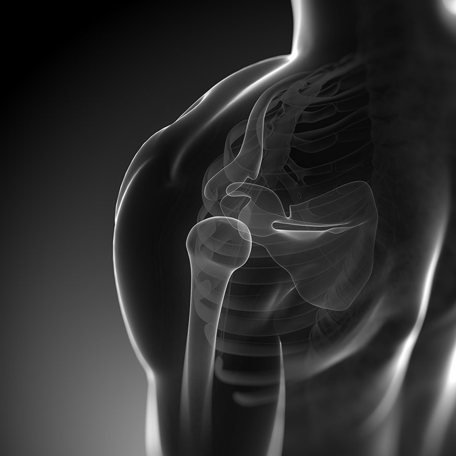
SEPTEMBER 29, 2016
Was tun bei Verletzungen?
Klassische Risikofaktoren sind vor allem Selbstüberschätzung und falsche Ausrüstung – prüfen Sie also gerade zu Beginn einer neuen Saison den Zustand Ihrer Utensilien und steigern Sie sich beim Training langsam.
PECH-Regel: Pause – Eis – Kompression – Hochlagern.
Die am häufigsten von Sportverletzungen betroffenen Körperpartien sind das Knie, das Sprunggelenk und die Schulter. Direkt nach der Verletzung gilt die PECH-Regel: Pause / Eis, also Kühlung der betroffenen Stelle / Kompression, also das Anlegen eines festen Verbandes / Hochlagern der Extremität. Das „return to sports“, die Zeit, die der Körper zur Regeneration und für die Rückkehr zur alten Form benötigt, ist nicht nur stark abhängig von der Erstversorgung, sondern auch von der unmittelbaren Weiterbehandlung und Physiotherapie.
Wie können
wir helfen?
Bei Verletzungen, Arbeits- und Wegeunfällen stehen wir Ihnen ab der Notfallversorgung durchgehend zur Seite, von der Therapie bis zur Rehabilitation: So ermöglichen wir beste Heilungschancen bei kurzen Ausfallzeiten.
So erreichen Sie uns:
